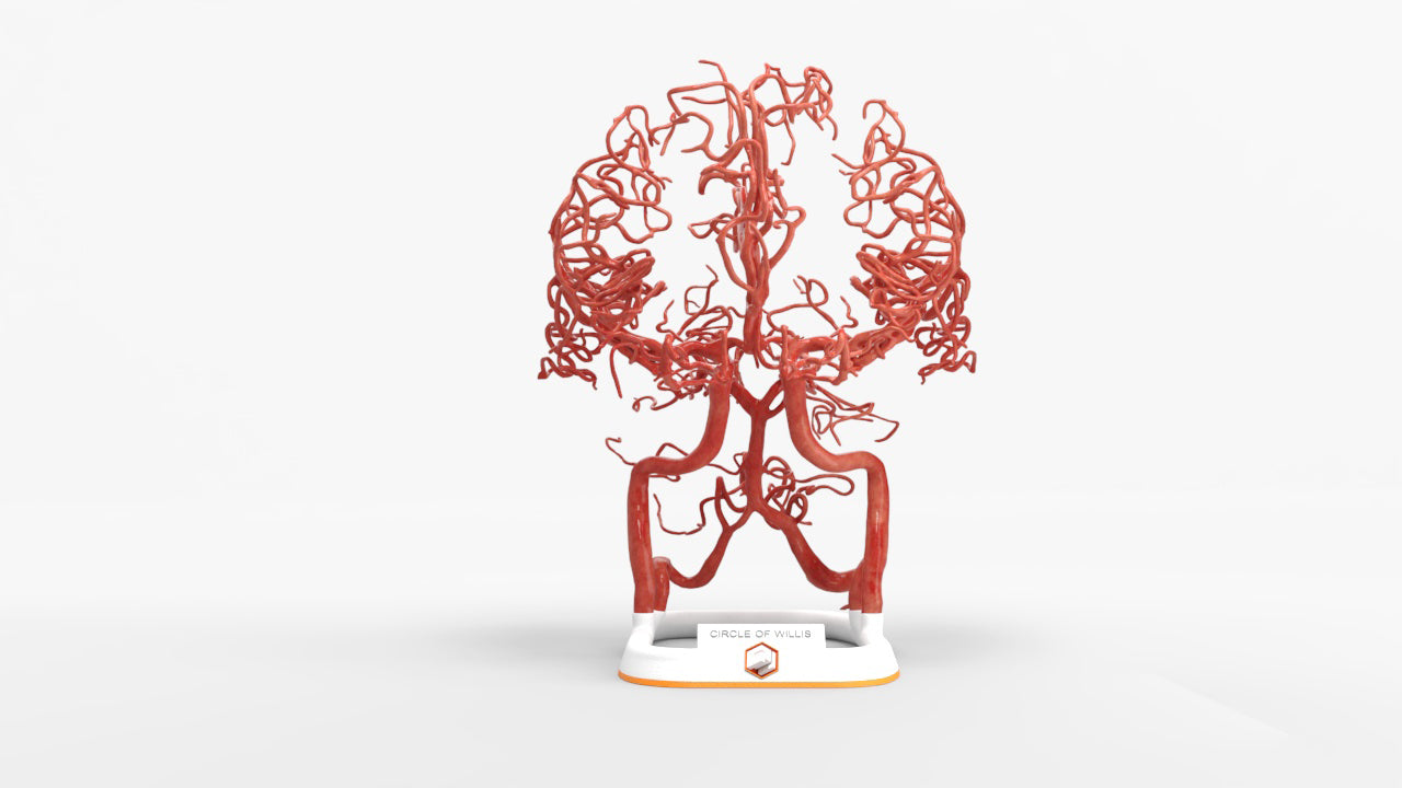
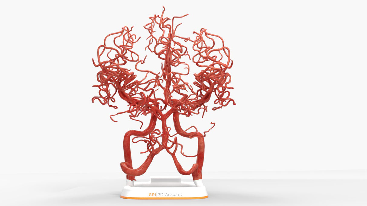
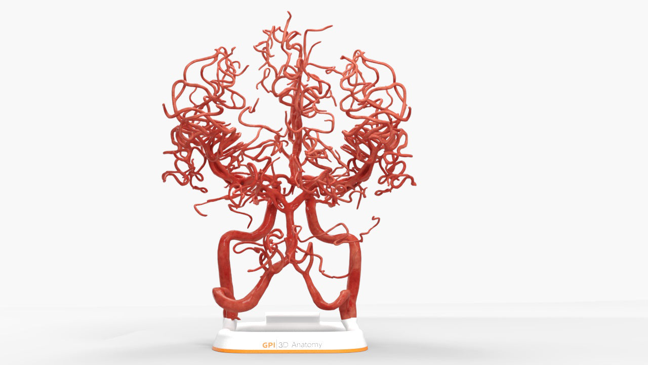
Circle of Willis (Circulus Arteriosus Cerebri) - Male, 54

$1,325.00
Shipping calculated at checkout.

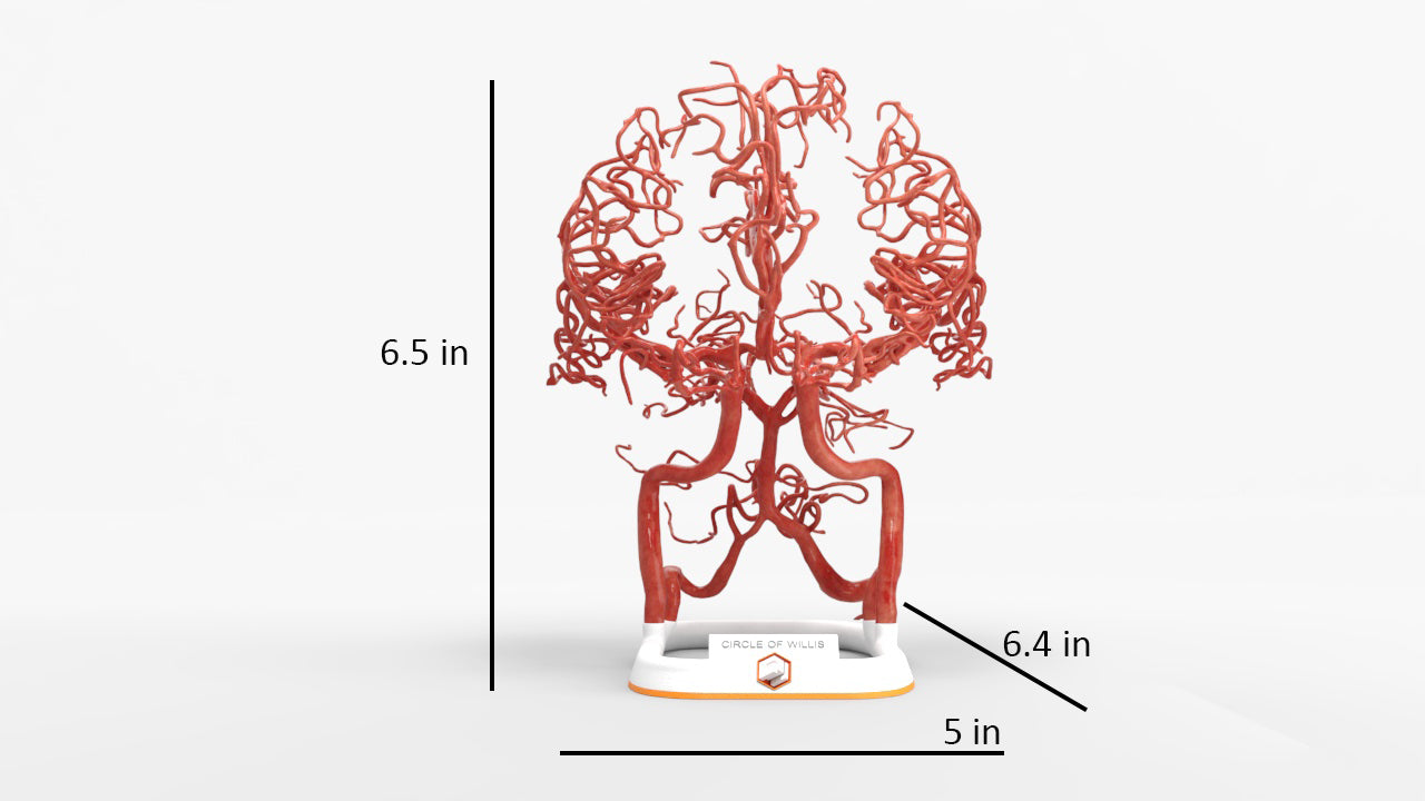
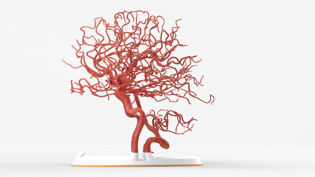
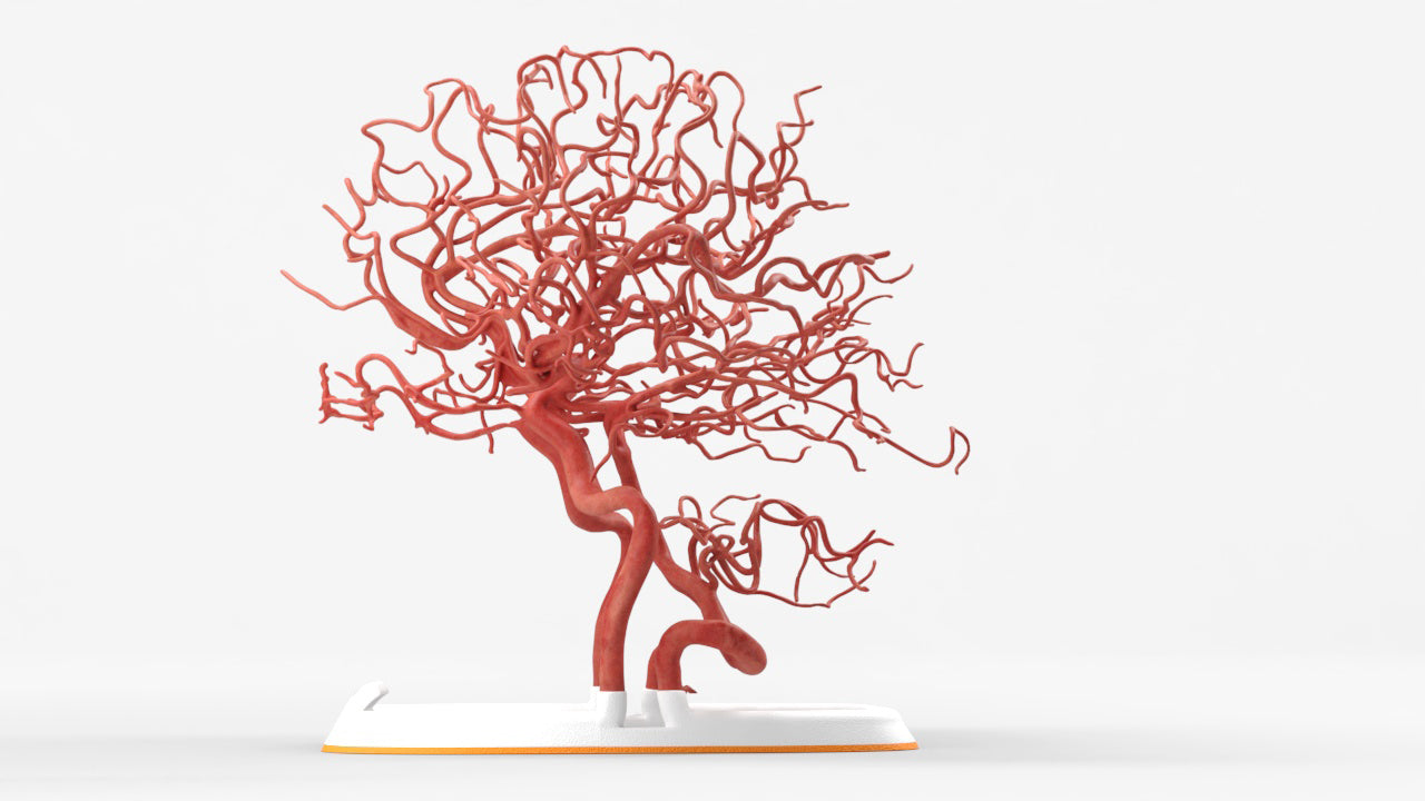
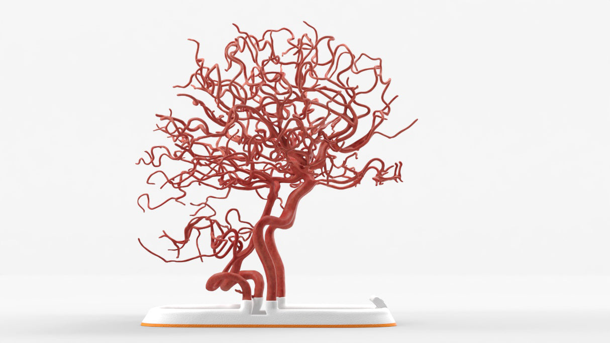
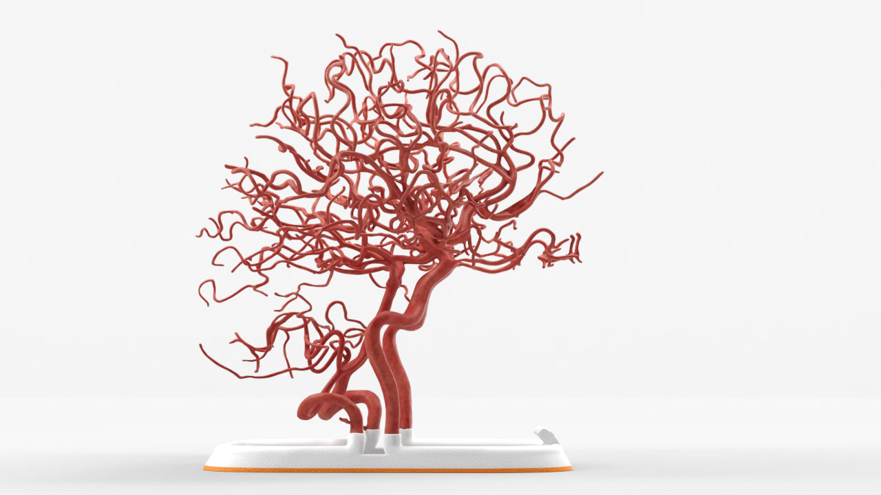
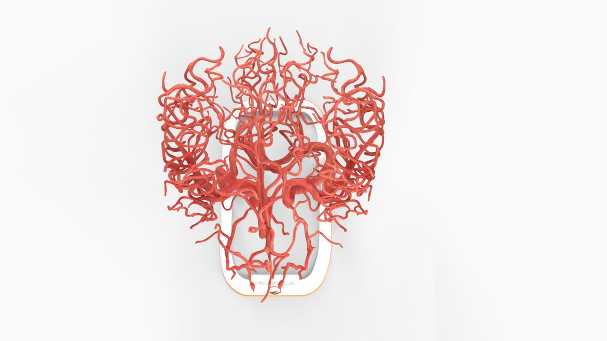
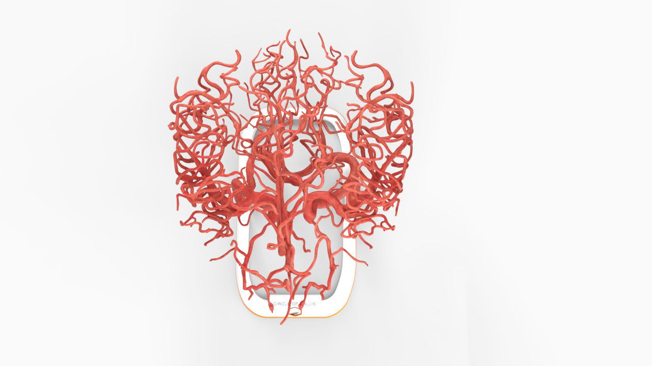
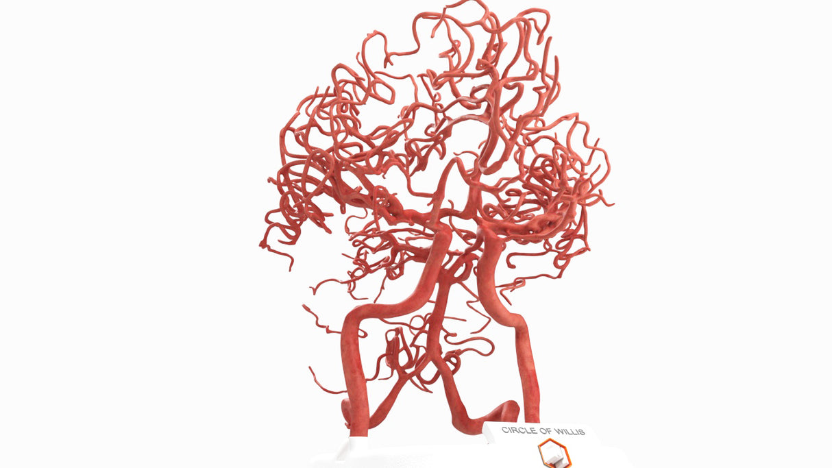
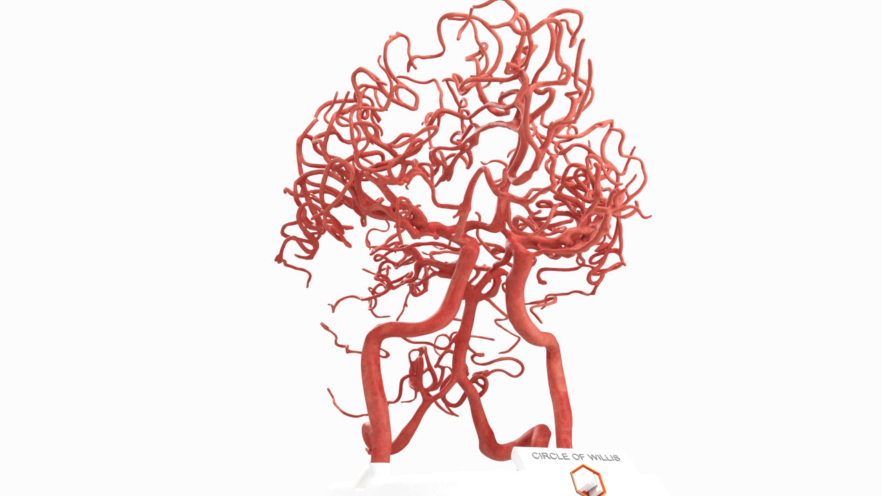
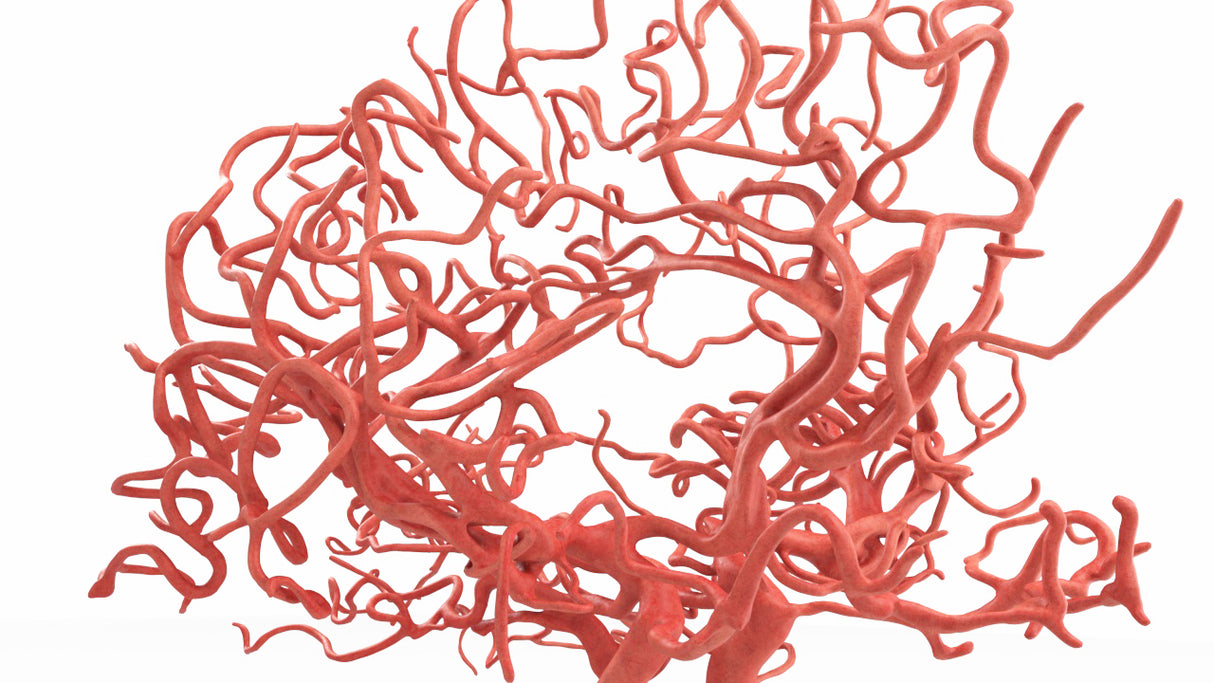
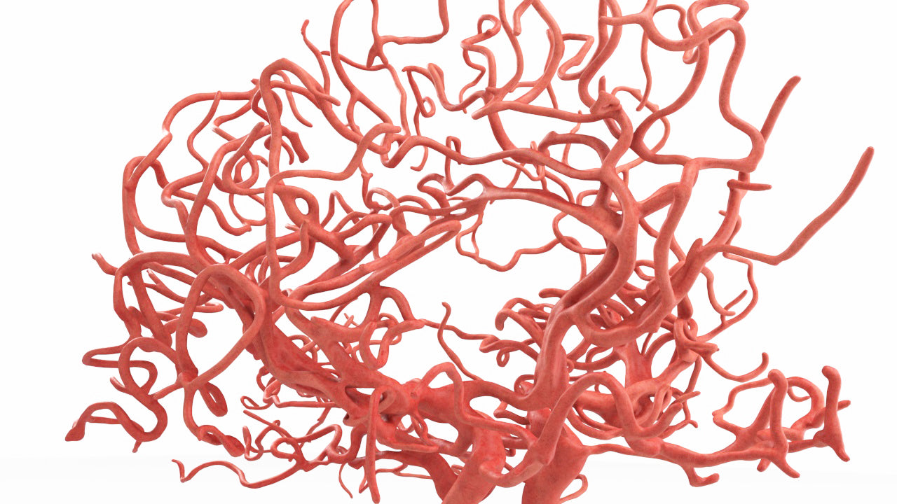
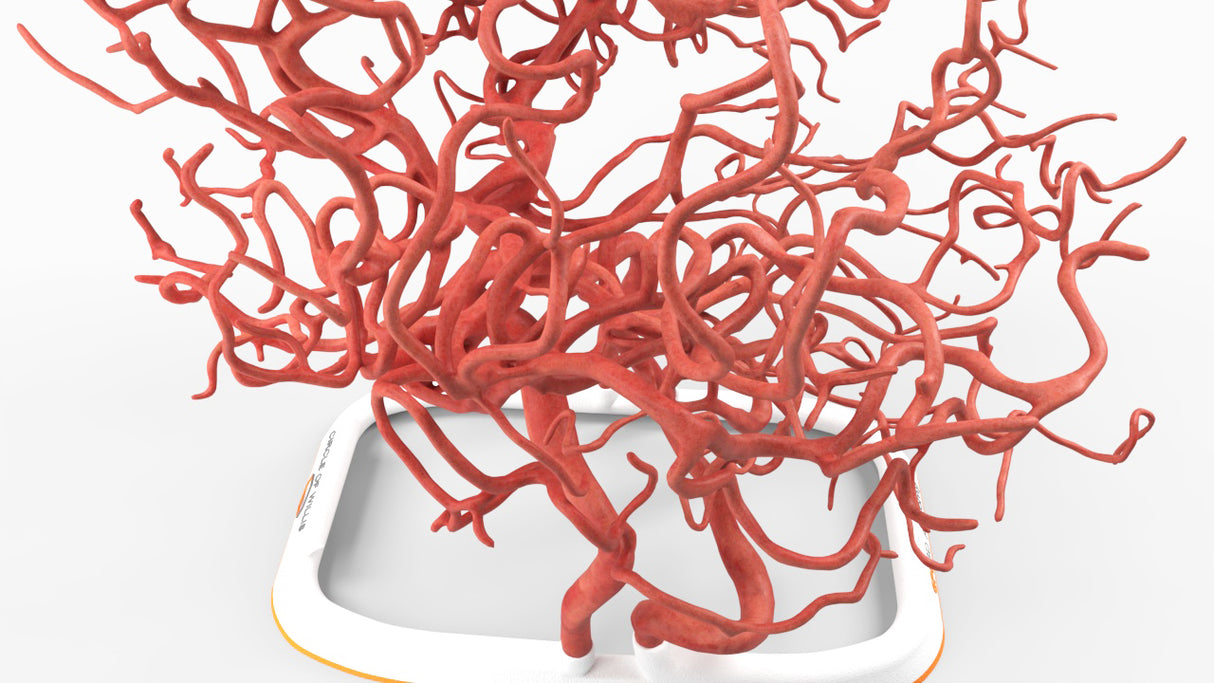
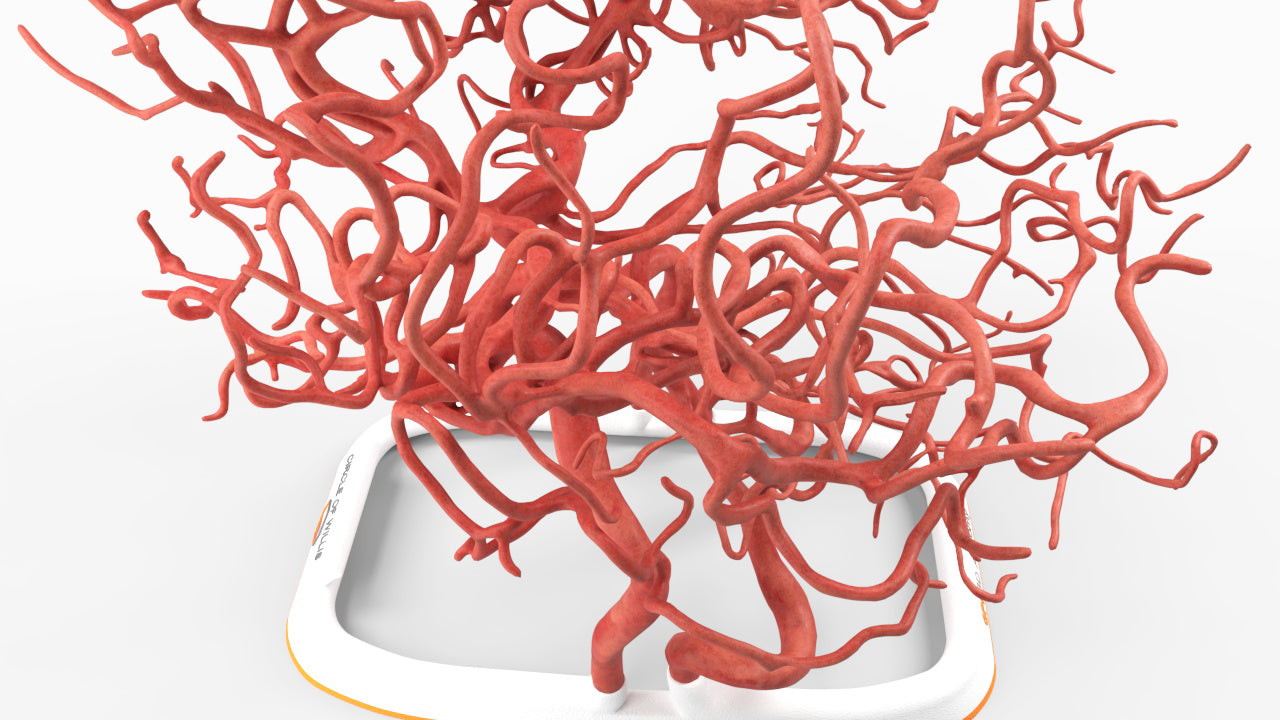
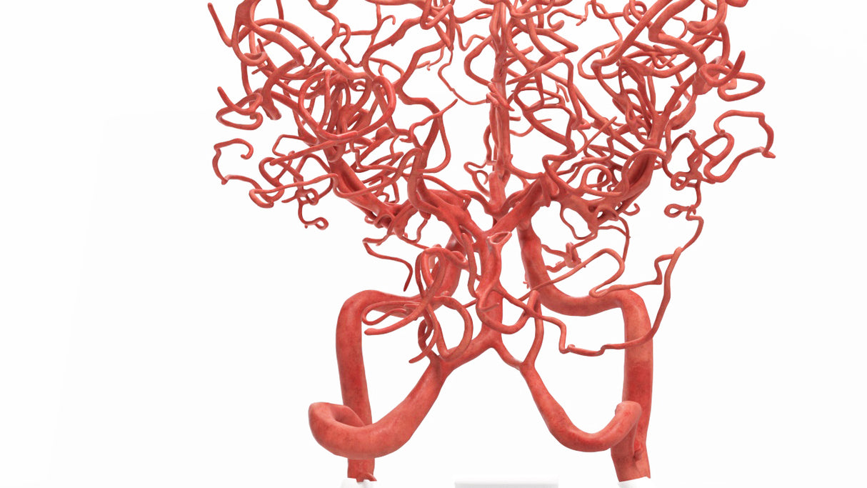
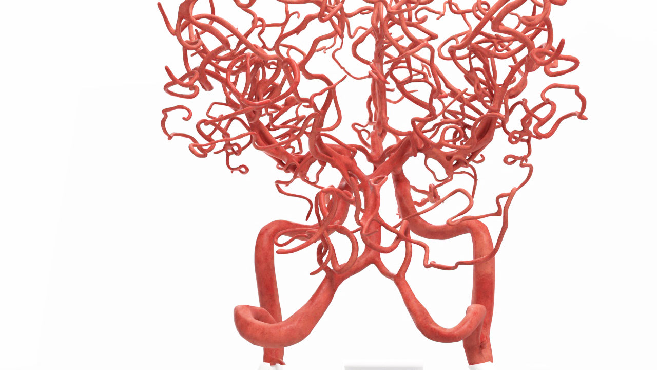
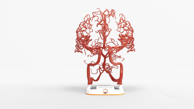
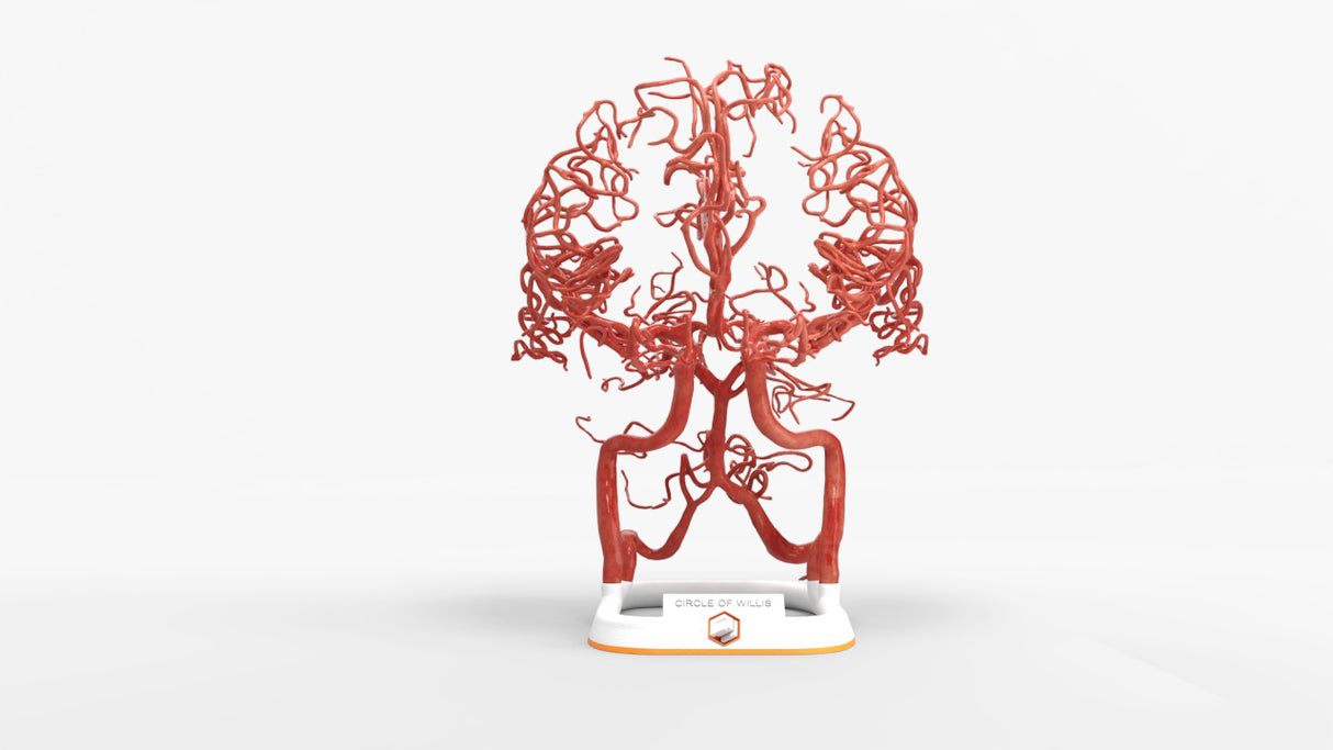
Circle of Willis (circulus ateriosus cerebri), an anastomosis between the anterior and the posterior communicating arteries. A symmetrical circle, found in less than half of brains, connects the two major arterial systems, the internal carotid arteries and the vertebrobasilar (vertebral and basilar arteries) systems. Artery size varies widely within the same structure and between individuals.  


Includes left and right internal carotid artery (ICA), anterior cerebral artery (ACA) and middle cerebral artery (MCA).

Designed using real patient imaging scans and the latest 3D printing technologies, in collaboration with Mayo Clinic.

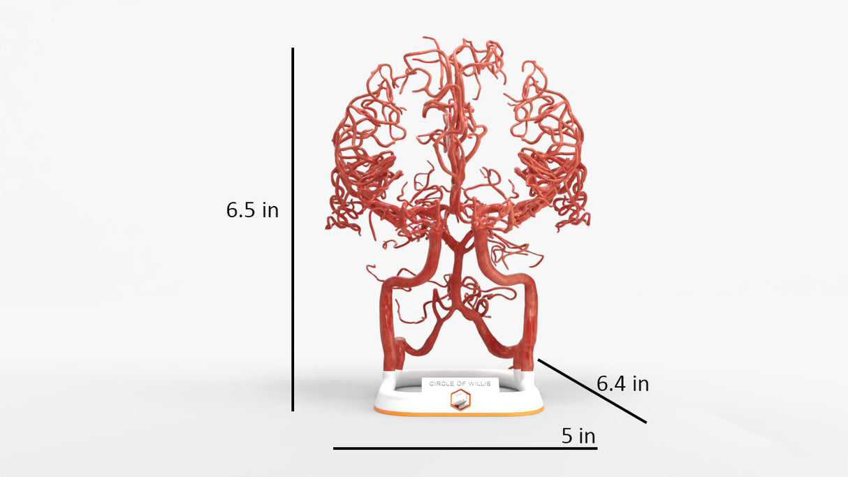
Dimensions & Features

100%: 6.4in x 5in x 6.5in

About the Condition

What Is Circle of Willis?

The circle of Willis is a ring of arteries located at the base of the brain that supplies the majority of blood flow to the brain. The circle of Willis surrounds the pituitary gland and enables communication between the front and back of the brain, as well as between the left and right hemispheres of the brain. 

The circular network of arteries in the Circle of Willis helps protect against damage from a stroke or aneurysm. If a blockage or narrowing occurs in one area, blood flow can be rerouted to other vessels. 

Structure

The circle of Willis starts where the internal carotid arteries enter the brain. It then branches into the cerebral and communicating arteries, which form a circle around the base of the brain. 

Anterior (Front) Arc

  • Anterior cerebral arteries: Supplies blood to the motor and sensory cortex that controls the lower limbs.
  • Anterior communicating artery: Connects the left and right anterior cerebral arteries and plays a role in vision.
  • Middle cerebral arteries: Supplies the motor and sensory cortex for the upper limb and face.

Posterior (Back) Arc 

  • Posterior cerebral arteries: Supplies blood to the occipital (visual) cortex.
  • Posterior communicating arteries: Connects an internal carotid artery to a posterior cerebral artery, a common place for an aneurysm which can lead to palsy.
  • Basilar artery: Supplies the medulla and the pons with blood.

Benefits of 3D Printing

3D-printed anatomy models offer a variety of advantages for surgical planning, patient education and medical research, including:

∙ Greater accuracy and detail than traditional anatomical models. 3D-printed models are created from digital scans of a patient's anatomy, which ensures that they are as close as possible to an exact replica of real human anatomy.

∙ More versatility than traditional anatomical models. 3D-printed models can be customized to meet your specific needs, whether planning a complex surgical procedure, training with real patient data or facilitating personalized patient communication.

Not limited to standard manufacturing, 3DP provides the best opportunity to produce accurate models in natural organic shapes, sizes, and colors; creating the best representation of real human anatomy.

Why Buy With Us

  • All models are designed and produced in collaboration with Mayo Clinic 3D Anatomical Modeling Laboratories.

  • GPI Anatomicals is a leading provider of anatomical models for medical education and research, with a 40-year history of innovation and its products are used by healthcare professionals around the world.

  • READY TO SHIP  IN 1 – 2 WEEKS

    Free Shipping – Ground

    Shipping Insurance

    A Signature will be REQUIRED at time of delivery

Product comparison grid

A table comparing the facets of 3 products
Facet
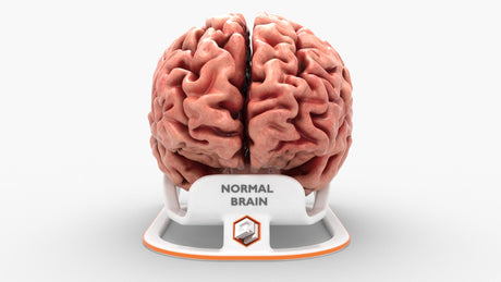
Adult Brain, Normal
View details
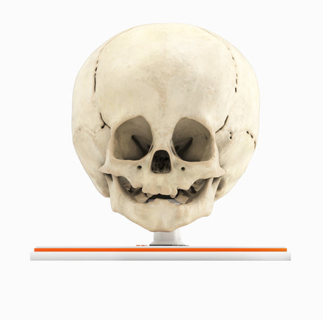
Infant Skull With Craniosynostosis of the Metopic Suture - Male, 8 Months
View details
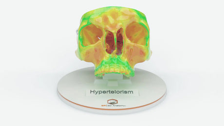
Hypertelorism – Male, 17 Years
View details
GPI 3D Anatomy

Circle of Willis (Circulus Arteriosus Cerebri) - Male, 54

$1,325.00

Circle of Willis (circulus ateriosus cerebri), an anastomosis between the anterior and the posterior communicating arteries. A symmetrical circle, found in less than half of brains, connects the two major arterial systems, the internal carotid arteries and the vertebrobasilar (vertebral and basilar arteries) systems. Artery size varies widely within the same structure and between individuals.  


Includes left and right internal carotid artery (ICA), anterior cerebral artery (ACA) and middle cerebral artery (MCA).

View product