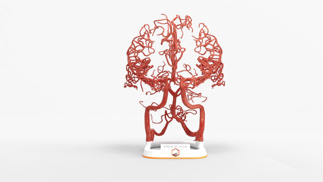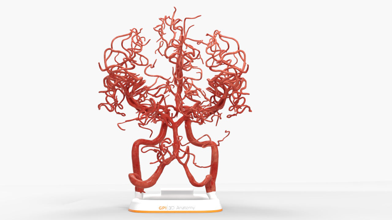
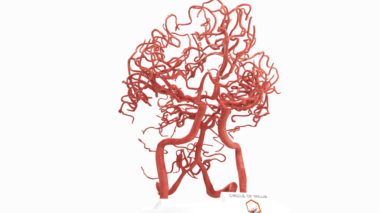
Circle of Willis (Circulus Arteriosus Cerebri) - Male, 54
Couldn't load pickup availability
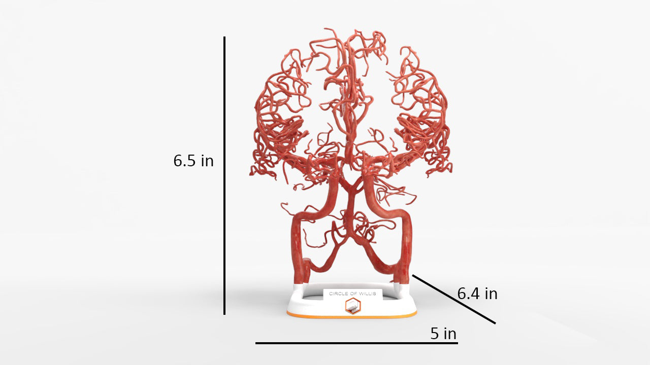
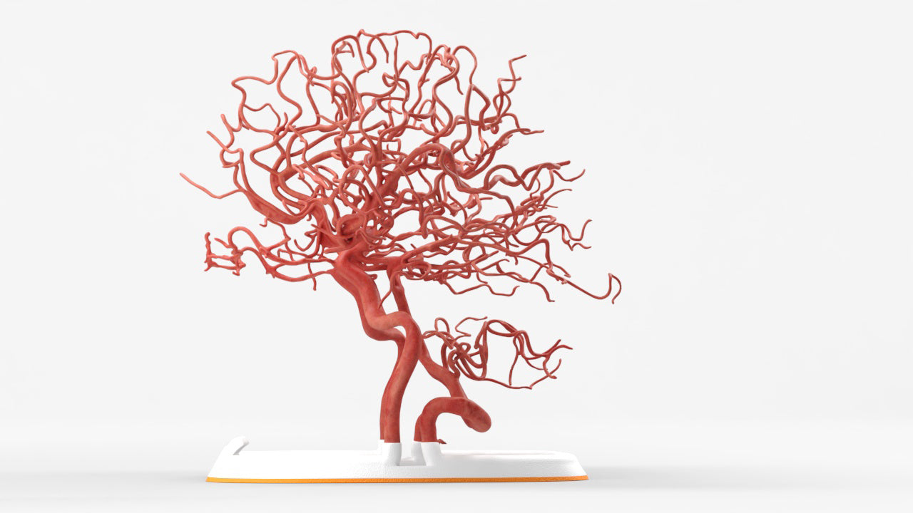
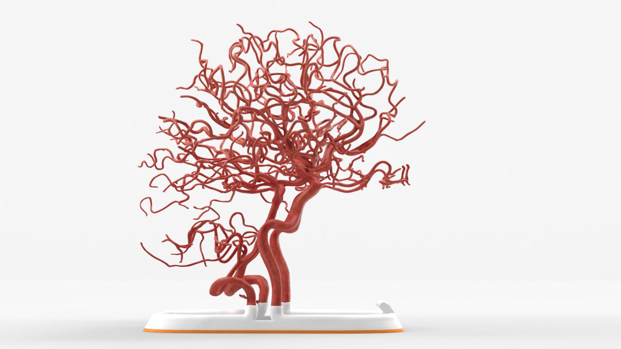
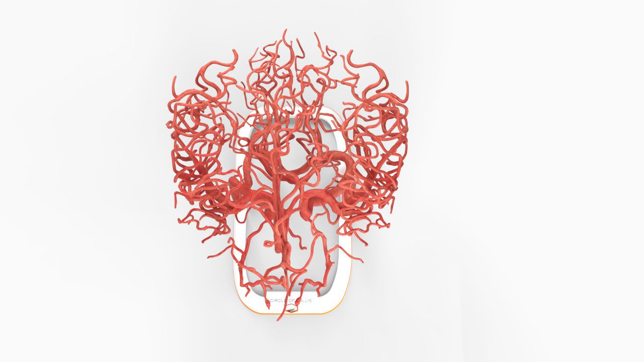
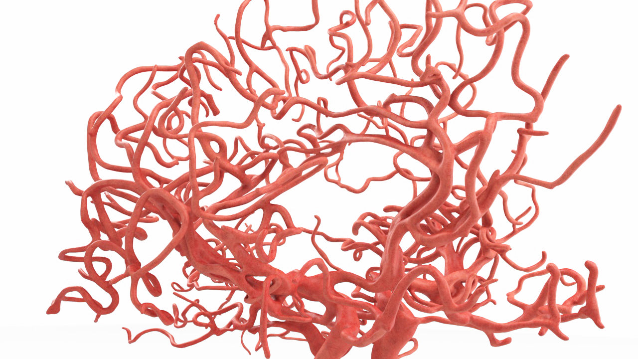
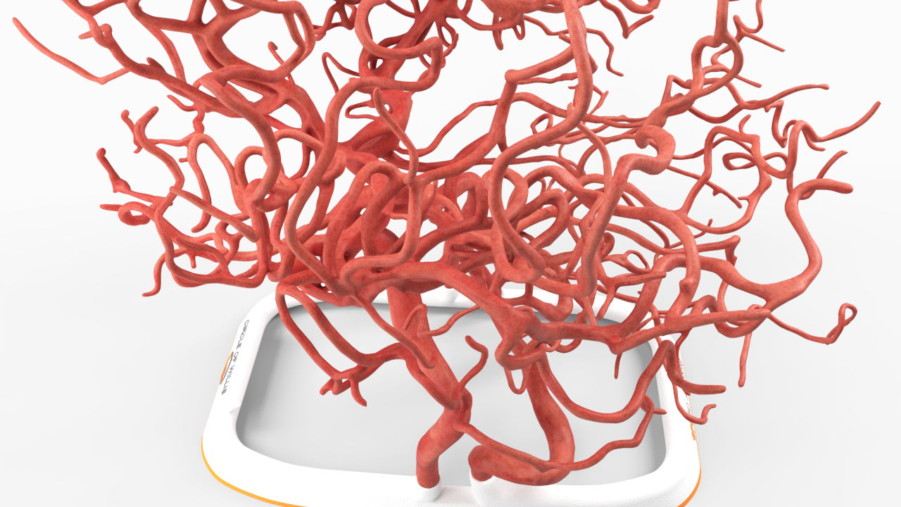
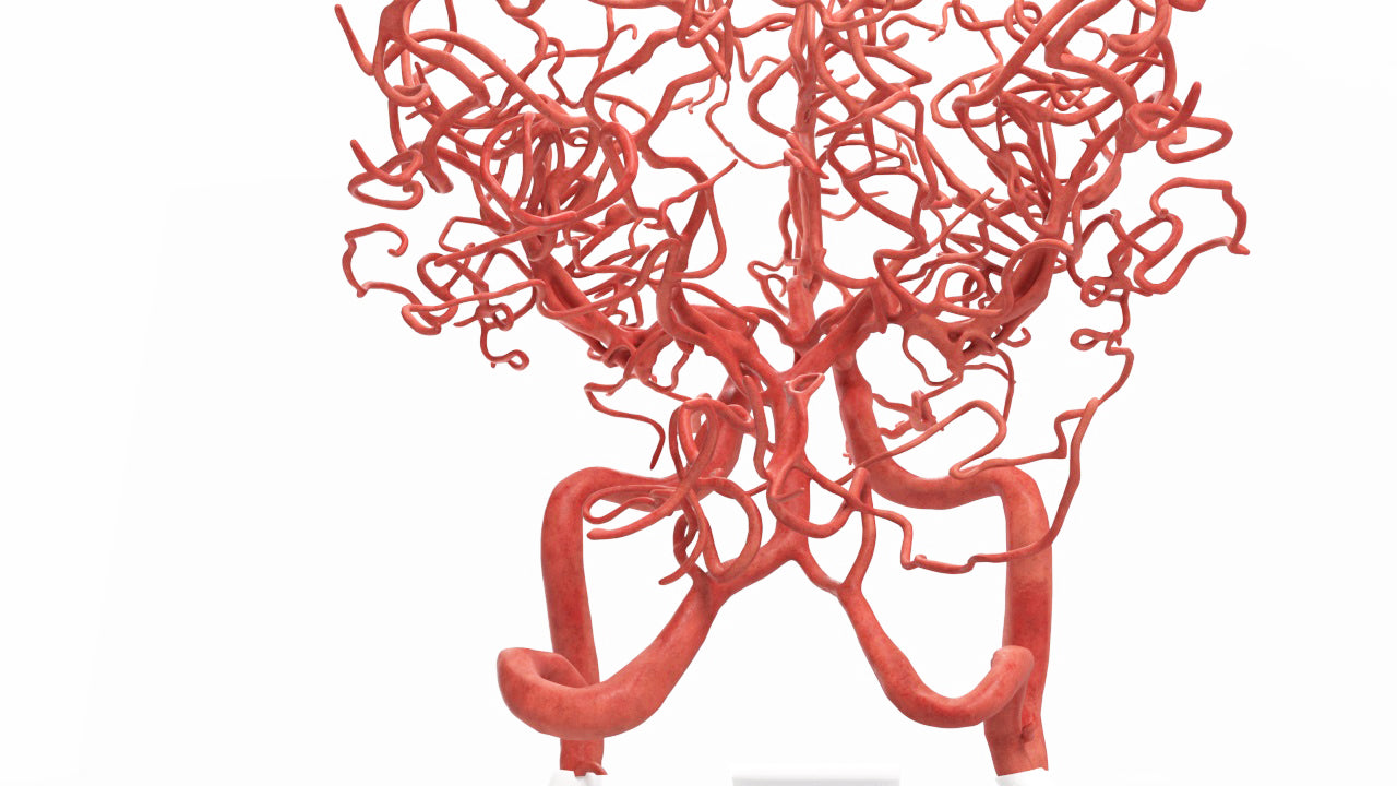
Circle of Willis (circulus ateriosus cerebri), an anastomosis between the anterior and the posterior communicating arteries. A symmetrical circle, found in less than half of brains, connects the two major arterial systems, the internal carotid arteries and the vertebrobasilar (vertebral and basilar arteries) systems. Artery size varies widely within the same structure and between individuals.
Includes left and right internal carotid artery (ICA), anterior cerebral artery (ACA) and middle cerebral artery (MCA).
Designed using MRI and CT imaging scans and the latest 3D printing technologies, in collaboration with Mayo Clinic.
Dimensions & Features

Circle of Willis (Circulus Arteriosus Cerebri) - Male, 54
About the Condition
Benefits of 3D Printing
3D-printed anatomy models offer a variety of advantages for surgical planning, patient education and medical research, including:
∙ Greater accuracy and detail than traditional anatomical models. 3D-printed models are created from digital scans of a patient's anatomy, which ensures that they are as close as possible to an exact replica of real human anatomy.
∙ More versatility than traditional anatomical models. 3D-printed models can be customized to meet your specific needs, whether planning a complex surgical procedure, training with real patient data or facilitating personalized patient communication.
Not limited to standard manufacturing, 3DP provides the best opportunity to produce accurate models in natural organic shapes, sizes, and colors; creating the best representation of real human anatomy.
Why Buy With Us
Product comparison grid
| Facet |
|---|