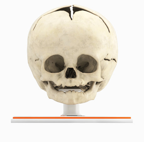
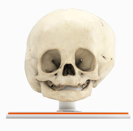
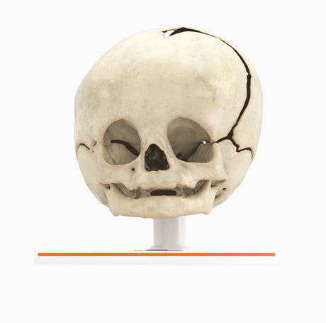
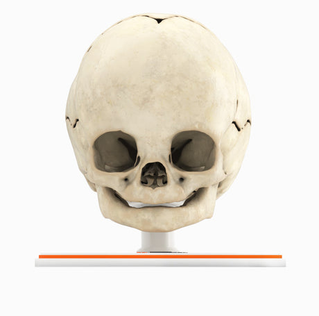
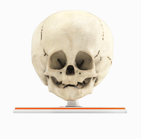
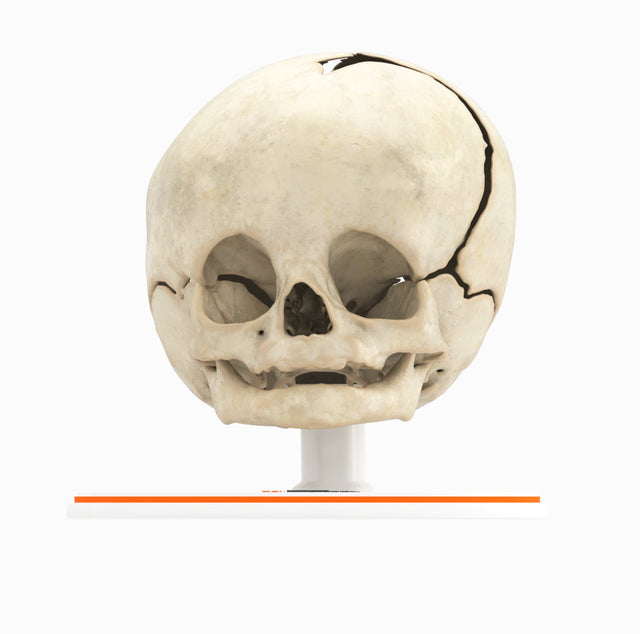
Infant Skull With Unilateral Craniosynostosis of the Coronal Suture - Male, 4 Months

Pricing and Availability Upon Request

Contact us: sales@gpianatomy.com


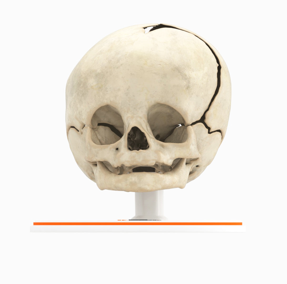
Infant Skull With Unilateral Craniosynostosis of the Coronal Suture - Male, 4 Months

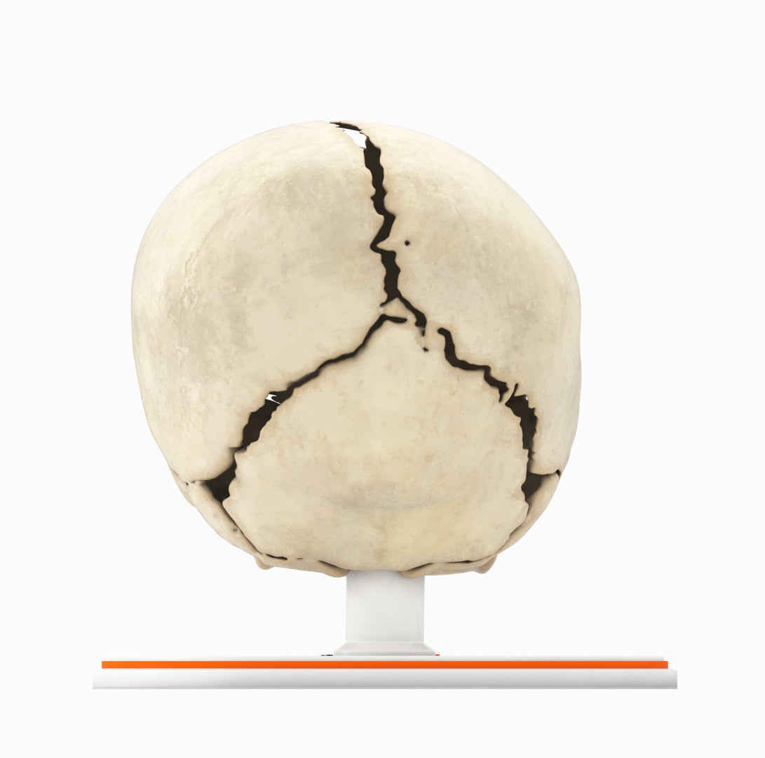
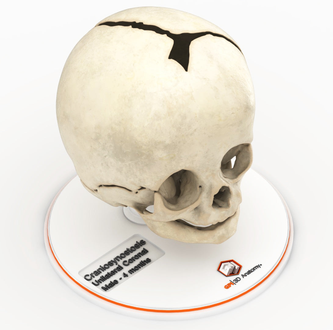
Craniosynostosis, a condition in which one or more cranial sutures in the infant skull prematurely mineralize and fuse before completion of brain development, with affected sutures: 

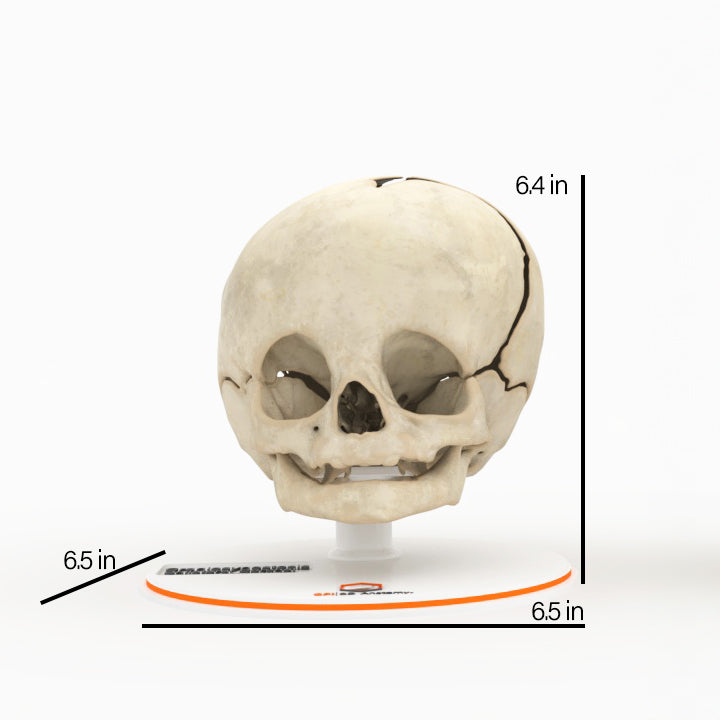
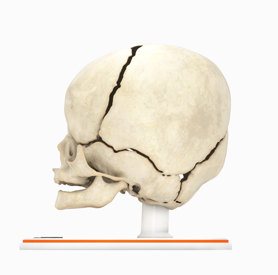
Unilateral coronal synostosis (anterior plagiocephaly) The coronal suture separates the two parietal bones from the frontal bone. Premature unilateral fusion flattens the forehead and brow on the affected side, with prominence on the unaffected side, giving the head a rotated shape.

In most cases, the cause of craniosynostosis is unknown. Crouzon, Apert and Pfeiffer syndromes are the most common craniofacial syndromes, accounting for nearly two-thirds of syndromic cases. Most of these patients exhibit elevated intracranial pressure (ICP), hydrocephalus, optic atrophy, and respiratory, speech and hearing problems. Surgical management is common for primary craniosynostosis where there is restriction of brain growth and elevated ICP, typically within the first year of life.

Designed using real patient imaging scans and the latest 3D printing technologies, in collaboration with Mayo Clinic.

Dimensions & Features

100%: 6.5in x 6.5in x 6.3in

About the Condition

What Is Craniosynostosis?

The bones of the skull are connected with fibrous joints called sutures. In between the sutures are membranous gaps called fontanelles, also known as soft spots. Sutures remain open to allow for bone expansion during fetal and childhood development. 

Craniosynostosis occurs when one or more of the sutures closes early. Premature fusion of sutures can cause the skull to grow in an unusual shape. Left untreated, craniosynostosis can restrict head and brain growth, and increase intracranial pressure. In addition to an abnormal skull shape, symptoms may include headache, projectile vomiting, fatigue, a bulging soft spot and difficulty looking upward. Left untreated, craniosynostosis can also interfere with psychosocial development and peer relationships later on.

Types of Cranial Sutures

Sutures are found throughout the surface of the skull. Some sutures run along the midline, but most are located on both sides of the skull as a pair.

The sagittal suture runs along the midline of the skull from front to back. Premature fusion causes a long, narrow skull. This is the most common type of synostosis.

The coronal suture runs across the top of the skull from ear to ear. Premature bilateral fusion can cause a short, wide skull. Premature unilateral fusion can flatten the forehead and brow on one side of the face, with a more prominent forehead and brow on the other side.

The metopic suture runs from the top of the skull down the middle of the forehead. Premature fusion gives the forehead a triangular shape and may affect the placement of the eyes. A metopic ridge is typically visible down the center of the forehead.

The lambdoid suture is located at the back of the skull. Premature fusion causes the skull to flatten in the back, pulling the forehead and ear with it, on one or both sides. It is the rarest type of synostosis.

How Common Is Craniosynostosis?

The condition occurs in one out of every 2,000 to 2,500 live births. It is more common in males than females by a ratio of 3:1. The cause of craniosynostosis is often unknown, but it can be related to certain genetic disorders including Apert, Crouzon and Pfeiffer Syndromes.

How Is It Treated?

Craniosynostosis may be treated a variety of traditional or minimally invasive surgical techniques, depending on the child’s age, affected sutures and other factors. Cranial molding helmets may be used alone or in conjunction with surgery. The goal of treatment is to correct the shape of the child’s head and allow for normal brain growth. Treatment is typically most effective as early as possible within the first year of an infant’s life.

Benefits of 3D Printing

3D-printed anatomy models offer a variety of advantages for surgical planning, patient education and medical research, including:

∙ Greater accuracy and detail than traditional anatomical models. 3D-printed models are created from digital scans of a patient's anatomy, which ensures that they are as close as possible to an exact replica of real human anatomy.

∙ More versatility than traditional anatomical models. 3D-printed models can be customized to meet your specific needs, whether planning a complex surgical procedure, training with real patient data or facilitating personalized patient communication.

Not limited to standard manufacturing, 3DP provides the best opportunity to produce accurate models in natural organic shapes, sizes, and colors; creating the best representation of real human anatomy.

Why Buy With Us

  • All models are designed and produced in collaboration with Mayo Clinic 3D Anatomical Modeling Laboratories.

  • GPI Anatomicals is a leading provider of anatomical models for medical education and research, with a 40-year history of innovation and its products are used by healthcare professionals around the world.

  • READY TO SHIP  IN 1 – 2 WEEKS

    Free Shipping – Ground

    Shipping Insurance

    A Signature will be REQUIRED at time of delivery

Product comparison grid

A table comparing the facets of 5 products
Facet
Infant Skull, Normal - Male, 8 Months
View details
Infant Skull With Unilateral Craniosynostosis of the Lambdoid Suture - Female, 7 Months
View details
Infant Skull With Unilateral Craniosynostosis of the Coronal Suture - Male, 4 Months
View details
Infant Skull With Craniosynostosis of the Sagittal Suture - Male, 8 Months
View details
Infant Skull With Craniosynostosis of the Metopic Suture - Male, 8 Months
View details
GPI 3D Anatomy

Infant Skull With Unilateral Craniosynostosis of the Coronal Suture - Male, 4 Months

$0.00

Infant Skull With Unilateral Craniosynostosis of the Coronal Suture - Male, 4 Months

Craniosynostosis, a condition in which one or more cranial sutures in the infant skull prematurely mineralize and fuse before completion of brain development, with affected sutures: 

Unilateral coronal synostosis (anterior plagiocephaly) The coronal suture separates the two parietal bones from the frontal bone. Premature unilateral fusion flattens the forehead and brow on the affected side, with prominence on the unaffected side, giving the head a rotated shape.

In most cases, the cause of craniosynostosis is unknown. Crouzon, Apert and Pfeiffer syndromes are the most common craniofacial syndromes, accounting for nearly two-thirds of syndromic cases. Most of these patients exhibit elevated intracranial pressure (ICP), hydrocephalus, optic atrophy, and respiratory, speech and hearing problems. Surgical management is common for primary craniosynostosis where there is restriction of brain growth and elevated ICP, typically within the first year of life.
View product