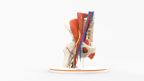
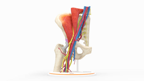
Hip Joint
The hip is the largest weight-bearing joint in the human body. A ball-and-socket joint, the hip joint consists of a ball (the top of the femur, also known as the thigh bone) and a socket (the acetabulum). Hip pain is most commonly due to degenerative arthritis and is a common orthopedic condition that may interfere with the ability to walk, stand, and, in severe cases, sleep.
Female Pelvic Floor
The female pelvic floor is composed of muscles that provide support to the pelvic organs (bladder, uterus, and rectum), aid in urinary and fecal continence, as well as contribute to sexual function and childbirth. The muscles of the pelvic floor are divided into two main layers:
1. Superficial Perineal Muscles
These muscles are located closer to the external genitalia and support the vaginal opening and include the bulbospongiosus, Ischiocavernosus, Superficial Transverse Perineal Muscle which stabilizes the perineal body, important for pelvic floor integrity, and external anal sphincter.
2. Deep Pelvic Floor Muscles or Pelvic Diaphragm
These muscles form the main support for the pelvic organs and include the levator ani group (Pubococcygeus, Puborectalis, and Iliococcygeus ), and the coccygeus (aka Ischiococcygeus)
Iliopsoas Muscles:
The psoas and iliacus muscles are collectively referred to as the iliopsoas complex. They are the dominant and strongest hip flexors, pulling the leg up at the hip joints. The psoas major originates from the lumbar spine, and the iliacus originates in the iliac fossa. Both merge inferiorly to form a common tendon inserting on the lesser trochanter of the femur. Tightness or weakness in this muscle can contribute to hip pain, lower back pain, and postural imbalances.
Lumbosacral Plexus:
The lumbosacral plexus is a network of nerves formed by the ventral rami of spinal nerves from L1 to S4. It provides motor and sensory innervation to the lower limb, pelvis, and perineum. The plexus is divided into two main parts:
Lumbar plexus (L1-L4): The femoral nerve, depicted in this model, is made from the L2-L4 nerves, providing motor function to the muscles of the quadriceps and sensation to the anterior thigh. It is intimately associated with the psoas muscles as it exits the spine and extends inferiorly between the psoas muscle and iliacus, exiting under the inguinal ligament just lateral to the iliopsoas tendon.
Sacral Plexus (L5-S4): The sacral plexus plays key roles in the motor, sensory, and autonomic functions of the pelvis, pelvic organs, perineum, and lower extremities.
Motor Function:
Pelvic Muscles:
Pudendal nerve (S2–S4): Innervates muscles of the perineum, including the external anal sphincter and external urethral sphincter, helping with urinary and fecal continence.
Nerve to levator ani (S3–S4): Supplies muscles that control the pelvic floor, contributing to pelvic support and continence.
Gluteal Muscles:
Superior gluteal nerve (L4–S1): Innervates the gluteus medius, gluteus minimus, and tensor fasciae latae, helping with hip abduction, medial rotation, and stabilizing the pelvis during walking.
Inferior gluteal nerve (L5–S2): Innervates the gluteus maximus, responsible for hip extension and lateral rotation of the thigh.
Sensory functions to the skin of the leg and foot:
Pelvic and Perineal Organs:
Pudendal nerve: Provides sensory innervation to the external genitalia and anus, and also contributes to the sensation of the perineum.
Autonomic Functions: The sacral plexus has important autonomic components that contribute to bladder control and sexual function:
Pudendal nerve: Controls the external anal sphincter and external urethral sphincter, important for voluntary control of urination and defecation.
Pelvic splanchnic nerves (from S2–S4): These parasympathetic fibers innervate the pelvic organs, such as the bladder (responsible for urinary bladder contraction) and the rectum (for defecation control).
Sciatic Nerve: L4-S3
The sciatic nerve is the largest and longest nerve in the body. It arises from the lumbosacral plexus (L4–S3) but has major contributions from L5 and S1, providing motor and sensory innervation to the posterior thigh, leg, and foot. It forms in the pelvis beneath the piriformis muscle in the gluteal region, exits the pelvis through the greater sciatic foramen, and travels down the posterior thigh, deep to the gluteus maximus. In the thigh, it innervates the hamstrings, allowing knee flexion and hip extension. In the lower leg, it supplies the gastrocnemius, soleus, anterior tibialis, and intrinsic muscles of the foot.