Canine Ear Model
Couldn't load pickup availability
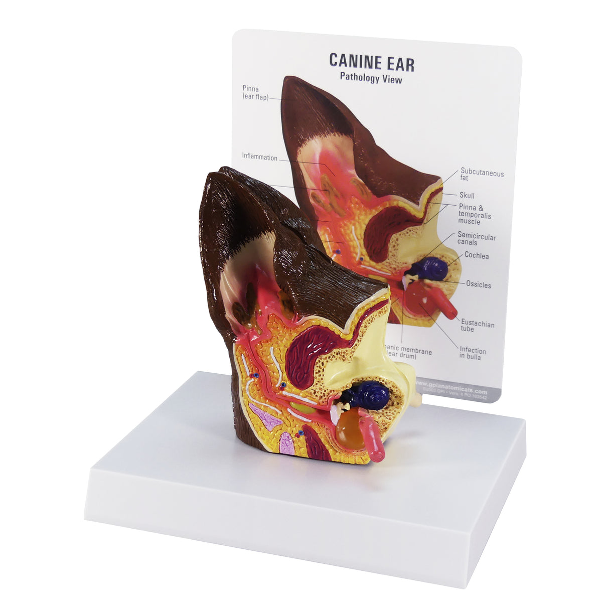
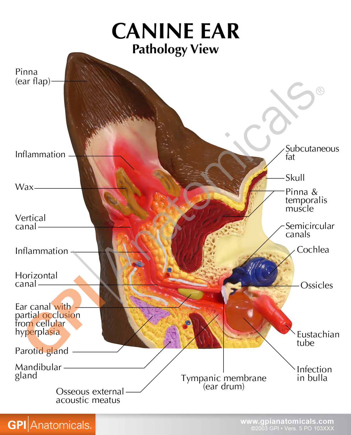
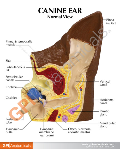
This two-sided, average size, canine ear model depicts: a normal side with cochlea, auditory ossicles, auditory tube, tympanic bulla, middle ear cavity, tympanic membrane, horizontal canal, vertical canal, auricular cartilage, pinna and temporalis muscle; abnormal side illustrates inflamed inner ear structures, inflammatory exudate in tympanic bulla, ear canal with partial occlusion from cellular hyperplasia, inflammatory exudate and an inflamed outer ear. This product contains a model, an informational card, & display base. The full size of the model measures 4-3/4” x 2-3/4” x 2”. The size of the card measures 5-1/4” x 6-1/2”. The size of the base measures 5-1/2” x 6-1/2”. Our model is great for veterinarian offices and classroom settings.

