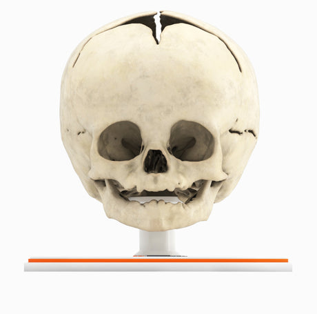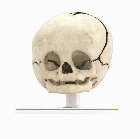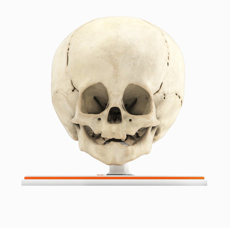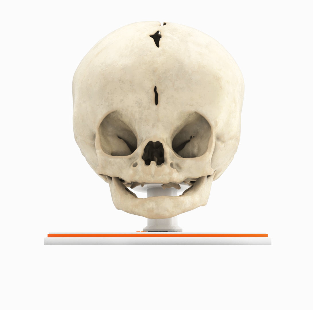
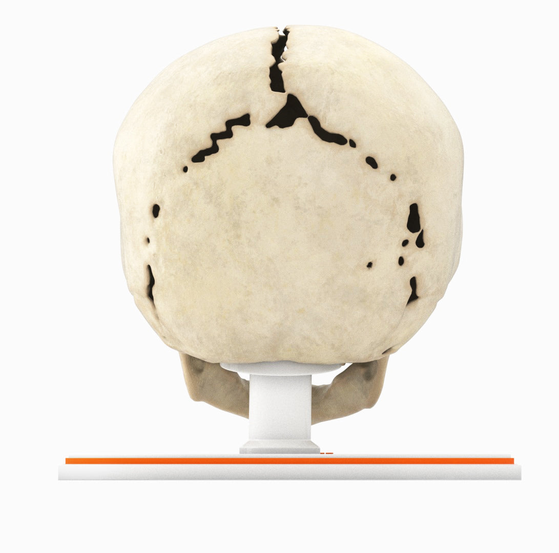

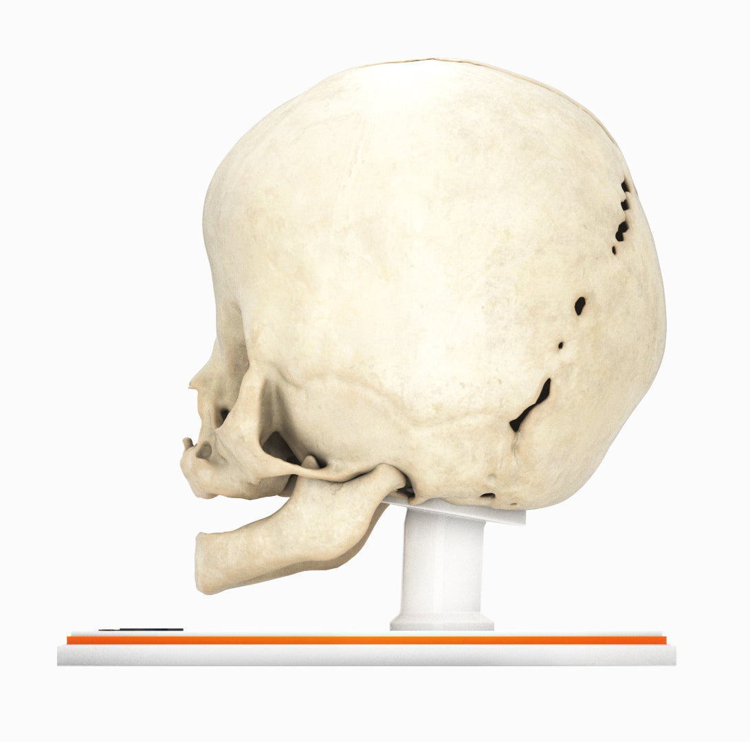
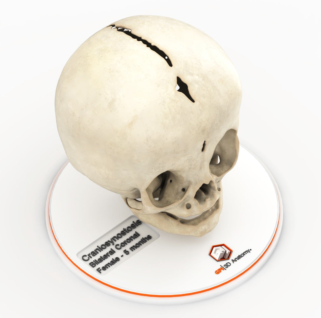
Infant Skull With Bilateral Craniosynostosis of the Coronal Suture - Female, 5 Months
Couldn't load pickup availability
Infant Skull With Bilateral Craniosynostosis of the Coronal Suture - Female, 5 Months
Craniosynostosis, a condition in which one or more cranial sutures in the infant skull prematurely mineralize and fuse before completion of brain development, with affected sutures:
Bilateral coronal synostosis (brachycephaly) – The coronal suture separates the two parietal bones from the frontal bone. Premature bilateral fusion flattens the forehead and brow, causing a short and wide skull and may cause vertical growth (turribrachycephaly).
In most cases, the cause of craniosynostosis is unknown. Crouzon, Apert and Pfeiffer syndromes are the most common craniofacial syndromes, accounting for nearly two-thirds of syndromic cases. Most of these patients exhibit elevated intracranial pressure (ICP), hydrocephalus, optic atrophy, and respiratory, speech and hearing problems. Surgical management is common for primary craniosynostosis where there is restriction of brain growth and elevated ICP, typically within the first year of life.
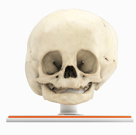
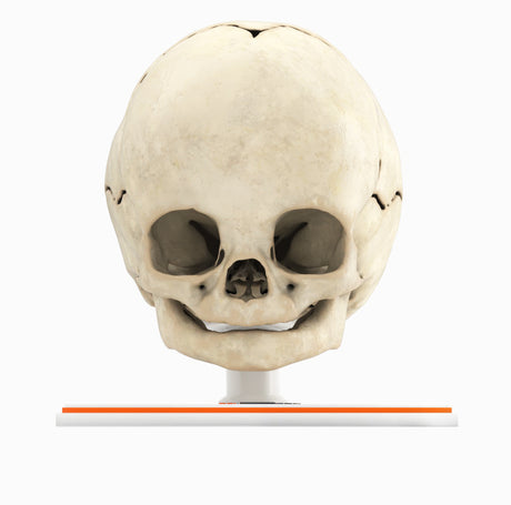
Designed using MRI and CT imaging scans and the latest 3D printing technologies, in collaboration with Mayo Clinic.
Dimensions & Features
Infant Skull With Bilateral Craniosynostosis of the Coronal Suture ...
About the Condition
Benefits of 3D Printing
3D-printed anatomy models offer a variety of advantages for surgical planning, patient education and medical research, including:
∙ Greater accuracy and detail than traditional anatomical models. 3D-printed models are created from digital scans of a patient's anatomy, which ensures that they are as close as possible to an exact replica of real human anatomy.
∙ More versatility than traditional anatomical models. 3D-printed models can be customized to meet your specific needs, whether planning a complex surgical procedure, training with real patient data or facilitating personalized patient communication.
Not limited to standard manufacturing, 3DP provides the best opportunity to produce accurate models in natural organic shapes, sizes, and colors; creating the best representation of real human anatomy.
Why Buy With Us
Product comparison grid
| Facet |
|---|