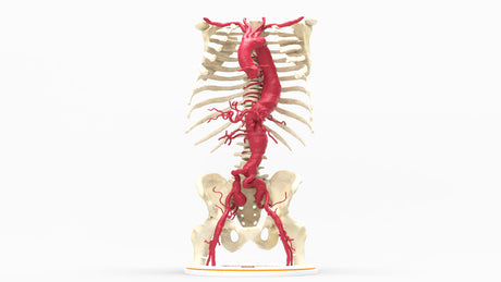
The Aorta
The aorta is the largest blood vessel in the body, serving as a conduit to deliver oxygenated blood to the head and body from the heart. In a resting state, it carries 5-6 liters of oxygenated blood per minute. The aorta is divided into four sections.
1. Ascending aorta: originates from the aortic valve proceeding upwards for approximately 5 cm. The ascending aorta starts at the aortic root and gives rise to the left and right coronary arteries, which supply oxygenated blood to the respective atriums and ventricles of the heart.
2. The aortic arch: curves to the left with several branches supplying blood to the head, neck, and upper extremities. It is the connection between the ascending and descending aorta with 3 main branches, including the brachiocephalic trunk, left common carotid artery, and left subclavian artery.
3. Descending aorta: Divided into two sections, thoracic and abdominal.
- Thoracic aorta is a continuation of the aortic arch supplying the bronchial arteries, which supply the lungs and bronchi, esophageal arteries, posterior intercostal arteries, and the superior phrenic arteries, which supply the diaphragm. As the thoracic aorta passes through the diaphragm via the aortic hiatus, it becomes the abdominal aorta, which supplies oxygenated blood to the abdominal organs and lower body.
- Abdominal aorta gives rise to the celiac trunk, which further branches into the left gastric artery supplying the stomach, the splenic artery supplying the spleen, and the common hepatic artery supplying the liver. The next major branch is the superior mesenteric artery (SMA), providing blood to the small intestine, ascending colon, and transverse colon. Paired renal arteries branch from the abdominal aorta to supply the kidneys, while the gonadal Arteries (testicular or ovarian arteries) deliver blood to the reproductive organs. The Inferior Mesenteric Artery (IMA) arises near the lower portion of the abdominal aorta, ensuring blood flow to the descending colon, sigmoid colon, and rectum.
4. Aortic bifurcation: The aorta terminates at L4, bifurcating into the right and left common iliac arteries. Each common iliac artery further branches into the Internal Iliac Artery, which supplies the pelvic organs, and the External Iliac Artery, which continues into the lower limb as the Femoral Artery, providing circulation to the legs and feet.
Aortic Wall:
The aorta consists of three layers of tissue:
• Tunica Intima (Inner Layer): Made of simple squamous endothelial cells that form a smooth lining, preventing friction of blood flow. Contains an internal elastic lamina, a thin layer of elastic fibers
• Tunica Media (Middle Layer): Thickest layer of the aortic wall composed of multiple layers of smooth muscle, and elastic tissues that allow the aorta to expand during systole and recoil during diastole. This layer assists in regulating blood pressure through controlled vasoconstriction and vasodilation.
• Tunica Externa (Outer Layer): This layer is composed of collagen fibers and connective tissues, which anchor the aorta to body tissue. It contains the vasum vasorum, a small blood vessel that nourishes the outer aortic wall.
What Conditions Affect the Aorta?
Several conditions can affect the aorta, potentially leading to serious complications.
• Aortic Aneurysm: A weakened area of the aortic wall that bulges outward, increasing the risk of rupture. It can occur in the ascending aorta, aortic arch, descending thoracic, or abdominal aorta.
• Aortic Dissection: A life-threatening condition where a tear forms in the inner layer of the aorta, causing blood to flow between the layers and potentially leading to rupture.
• Aortic Stenosis: Narrowing of the aortic valve, restricting blood flow from the heart to the aorta and forcing the heart to work harder.
• Aortic Regurgitation (Insufficiency): The aortic valve does not close properly, allowing blood to flow backward into the heart, leading to volume overload and heart strain.
• Coarctation of the Aorta: A congenital condition where a section of the aorta is abnormally narrowed, reducing blood flow to the lower body.
• Atherosclerosis of the Aorta: Buildup of fatty plaques in the aortic wall, which can narrow the artery, reduce blood flow, and increase the risk of aneurysms or dissections.
• Traumatic Aortic Injury: A rupture or tear in the aorta caused by severe chest trauma, often due to car accidents or falls.
• Congenital anomalies of the aorta, including right-sided aortic arch or duplicated aorta.