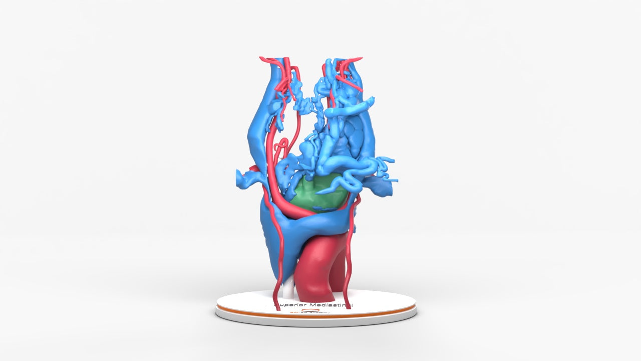
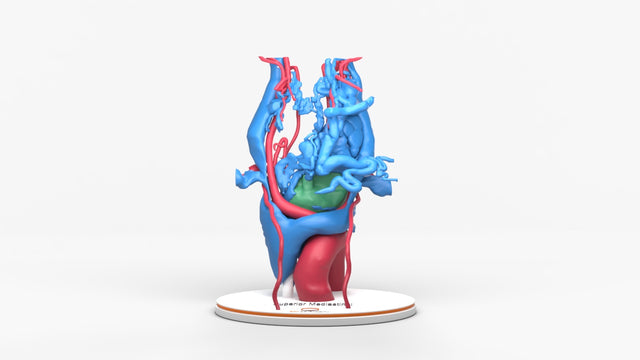
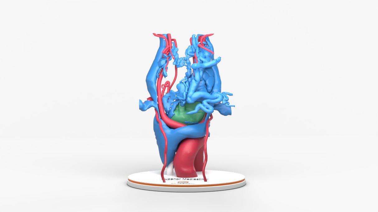
Chest With Mediastinal Paraganglioma – Male, 50 Years

Pricing and Availability Upon Request

Contact us: sales@gpianatomy.com


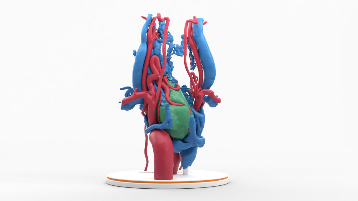
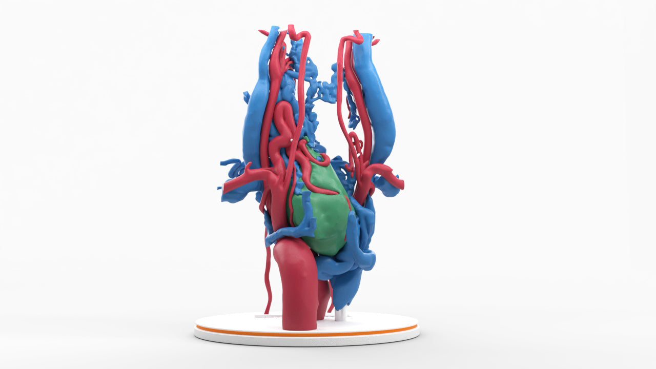
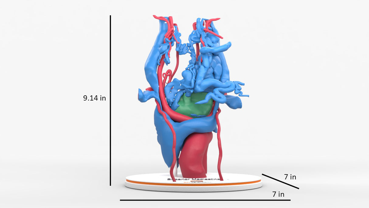
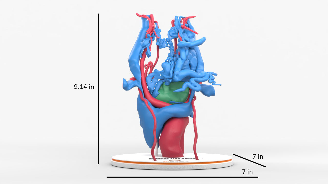
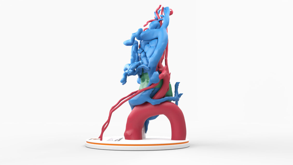
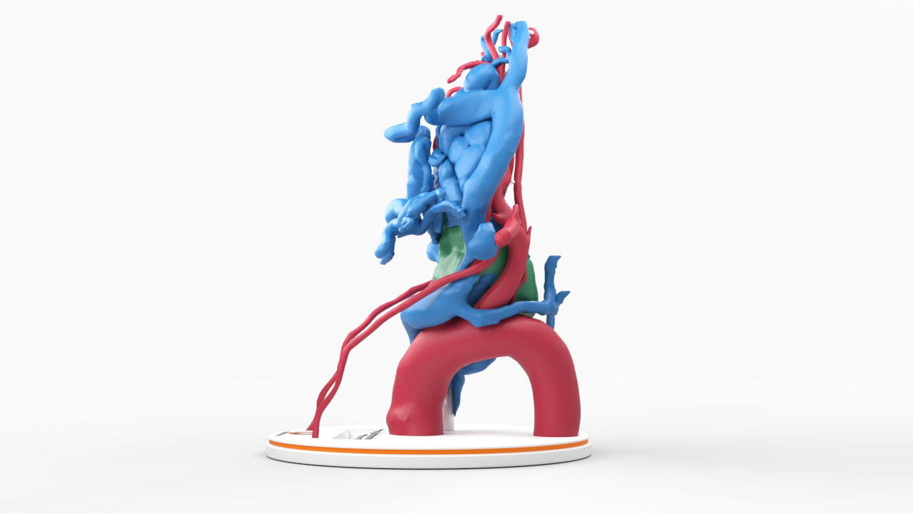
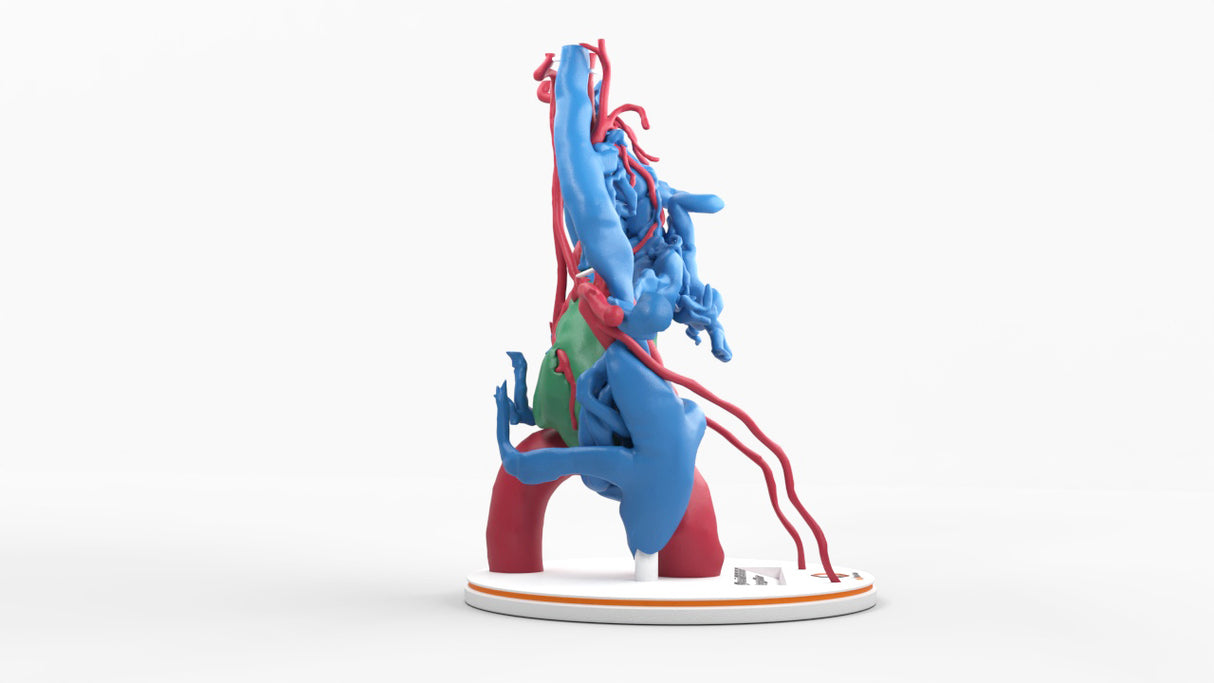
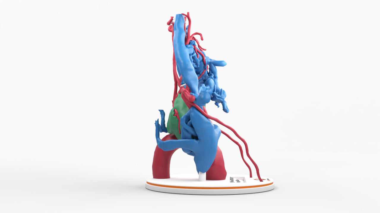
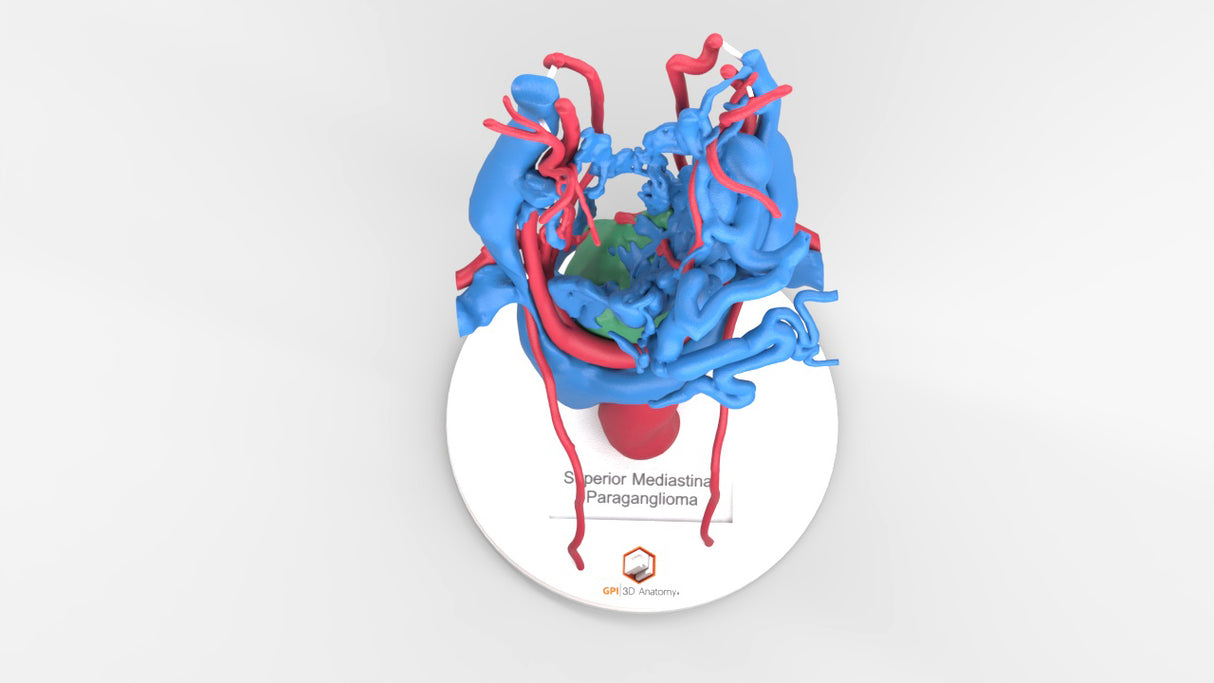
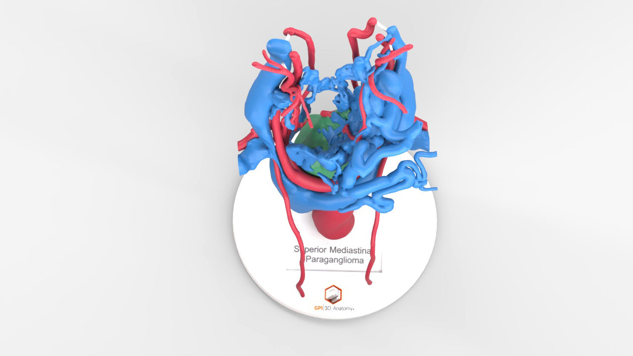
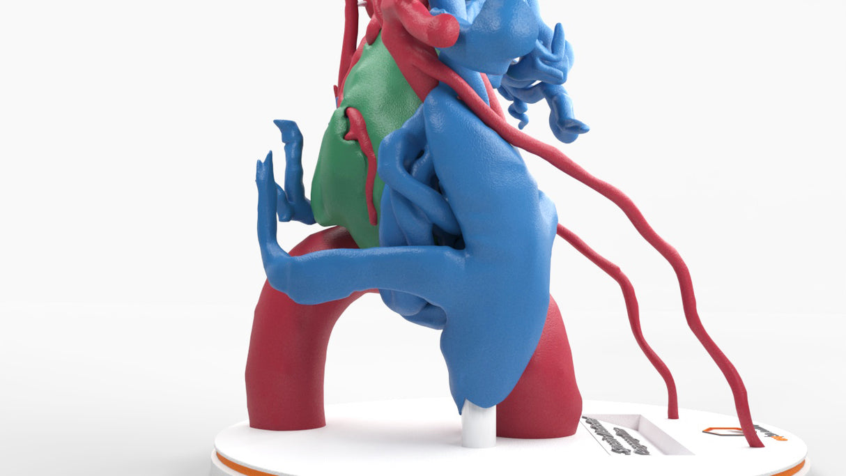
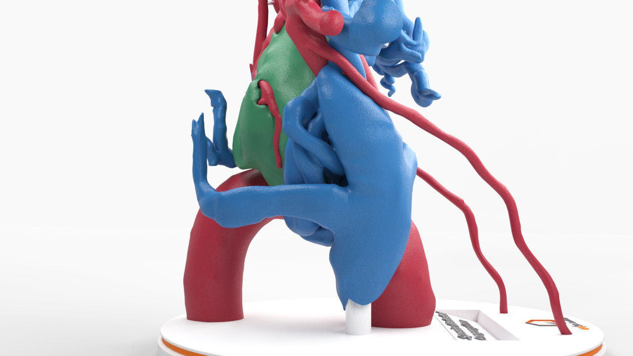
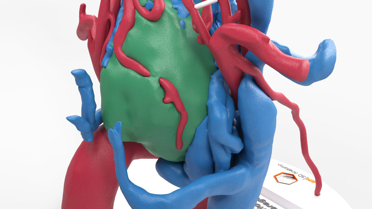
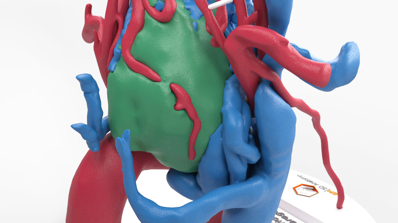
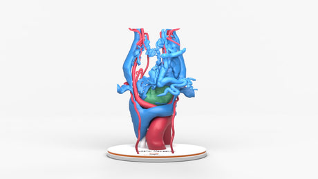
Chest with middle mediastinal paraganglioma, a rare tumor type derived from chromaffin cells (neuroectodermal cells), with abnormal increased arterial vascularity supplying the tumor and multiple enlarged draining veins. Due to the increased vascularity, this tumor type is at high risk of massive blood loss intraoperatively. Detailed surgical technique and preoperative blood pressure control are essential in the prevention and management of complications.

Complete surgical resection remains the standard of care. Prognosis after complete resection is favorable. 

Designed using real patient imaging scans and the latest 3D printing technologies, in collaboration with Mayo Clinic.

Dimensions & Features

100%: 7in x 7in x 9.4in

About the Condition

What Is a Paraganglioma?

Paragangliomas are rare neuroendocrine tumors. They can form in the chest and abdomen (called sympathetic) or the head and neck (called parasympathetic). The tumors are made of cells from the adrenal glands called chromaffin cells, which produce important hormones like dopamine, epinephrine and norepinephrine. Referred to as catecholamines, paragangliomas may release higher amounts of these hormones into the bloodstream. 

Most paranglioma tumors don’t cause symptoms. When they do, sympathetic paragangliomas may cause headaches, heart palpitations and high blood pressure. Parasympathetic paragangliomas may cause changes in vision, swallowing or ringing in the ears.

How Common Is a Paraganglioma?

The condition is extremely rare and occurs in roughly two out of every one million people. It is more common in females than males by a ratio of 3:1. It’s most likely to be diagnosed between the ages of 20 and 50, with 10% of cases occurring in children. Up to 40% of cases are caused by genetic conditions including: succinate dehydrogenase (SDH) mutations, RET mutations, NF1 mutations, von Hippel-Lindau syndrome and Carney-Stratakis syndrome/Carney triad. The cause is unknown in the remainder of cases.

How Is It Treated?

Most paragangliomas are benign when they’re diagnosed and successfully treated. Treatment typically includes surgery, followed by radiation therapy. In 25% of cases, the tumor is malignant and may spread to the lymph nodes, liver, lung or bones. Chemotherapy or other systemic treatment is often required. 

Benefits of 3D Printing

3D-printed anatomy models offer a variety of advantages for surgical planning, patient education and medical research, including:

∙ Greater accuracy and detail than traditional anatomical models. 3D-printed models are created from digital scans of a patient's anatomy, which ensures that they are as close as possible to an exact replica of real human anatomy.

∙ More versatility than traditional anatomical models. 3D-printed models can be customized to meet your specific needs, whether planning a complex surgical procedure, training with real patient data or facilitating personalized patient communication.

Not limited to standard manufacturing, 3DP provides the best opportunity to produce accurate models in natural organic shapes, sizes, and colors; creating the best representation of real human anatomy.

Why Buy With Us

  • All models are designed and produced in collaboration with Mayo Clinic 3D Anatomical Modeling Laboratories.

  • GPI Anatomicals is a leading provider of anatomical models for medical education and research, with a 40-year history of innovation and its products are used by healthcare professionals around the world.

  • READY TO SHIP  IN 1 – 2 WEEKS

    Free Shipping – Ground

    Shipping Insurance

    A Signature will be REQUIRED at time of delivery

Product comparison grid

A table comparing the facets of 3 products
Facet
Chest With Mediastinal Paraganglioma – Male, 50 Years
View details
Tracheobronchial Tree With Lymph Nodes - Male, 28 Years
View details
Chest Wall Tumor
View details
gpi-anatomicals-(trial)

Chest With Mediastinal Paraganglioma – Male, 50 Years

$0.00

Chest with middle mediastinal paraganglioma, a rare tumor type derived from chromaffin cells (neuroectodermal cells), with abnormal increased arterial vascularity supplying the tumor and multiple enlarged draining veins. Due to the increased vascularity, this tumor type is at high risk of massive blood loss intraoperatively. Detailed surgical technique and preoperative blood pressure control are essential in the prevention and management of complications.

Complete surgical resection remains the standard of care. Prognosis after complete resection is favorable. 

View product