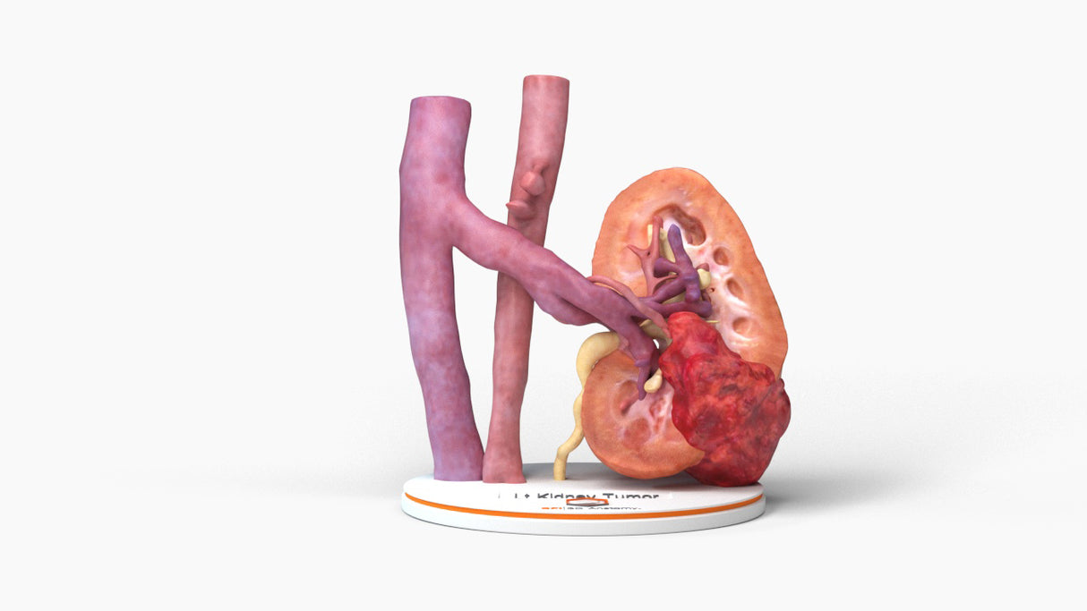
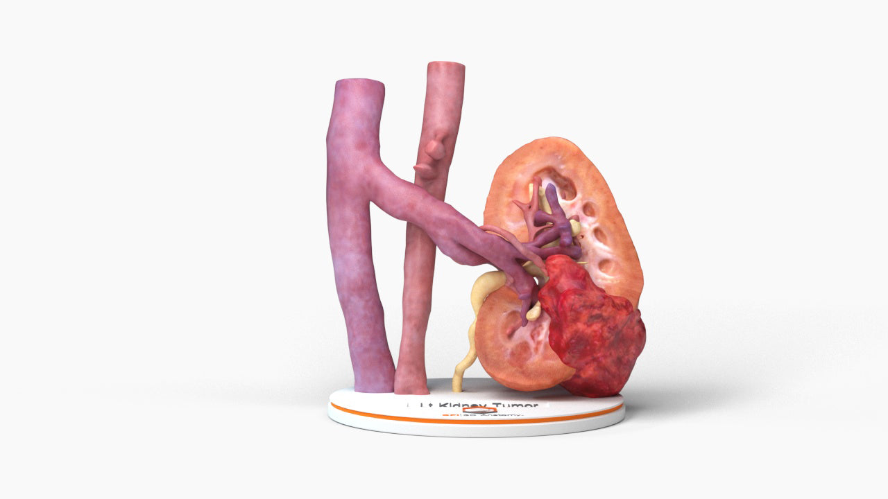
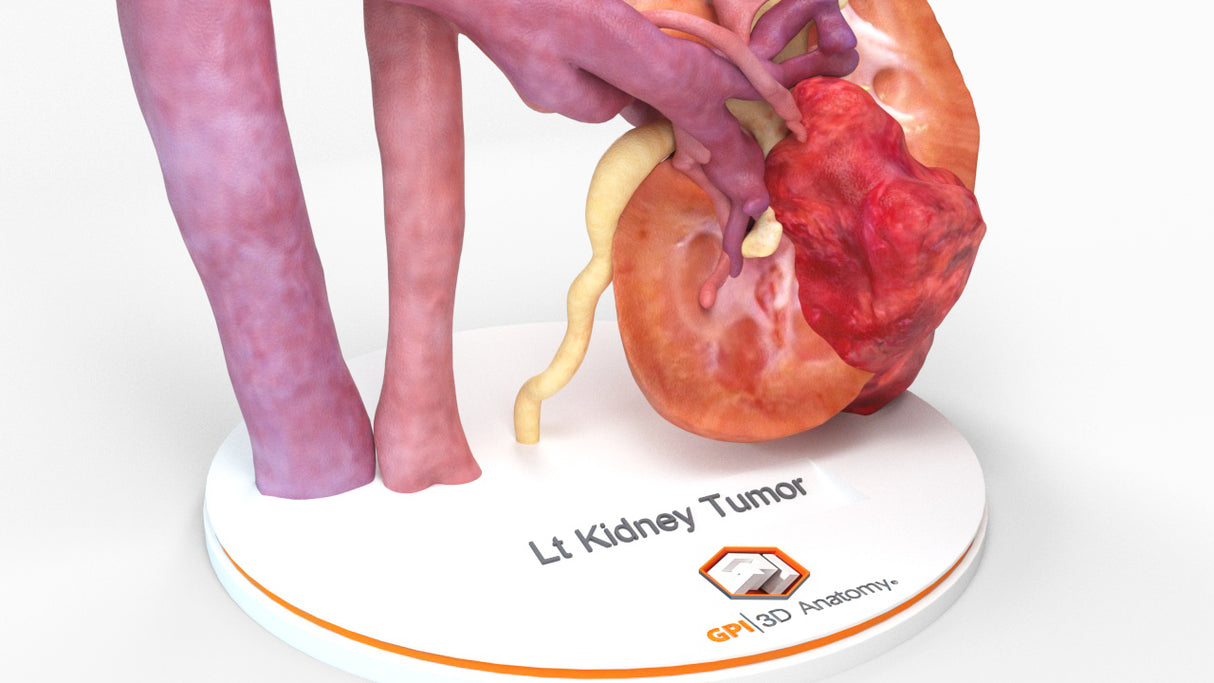
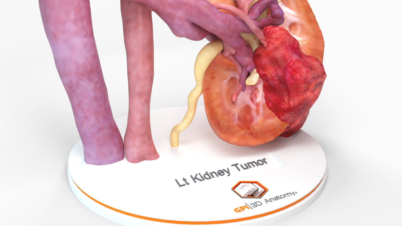
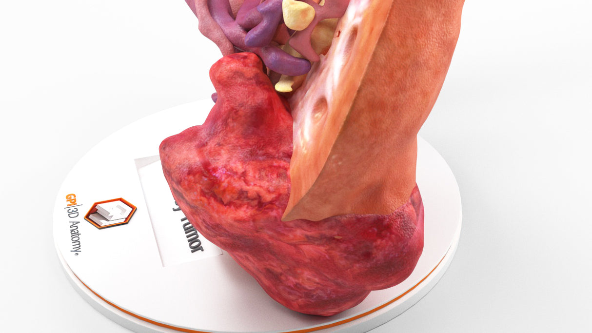
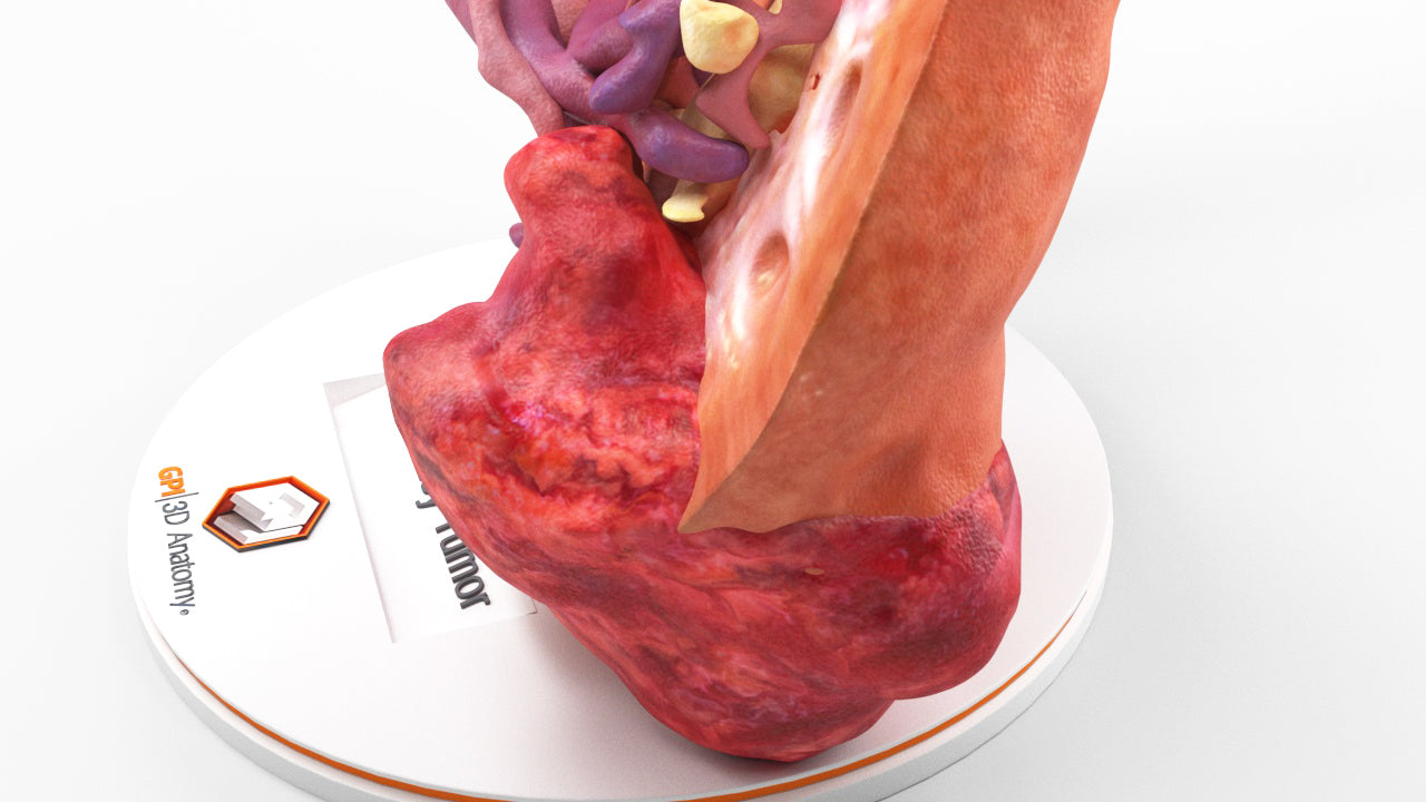
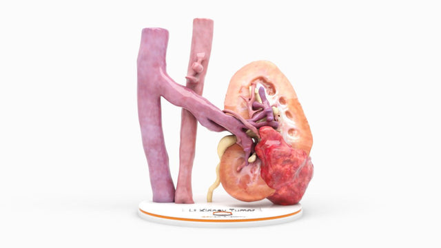
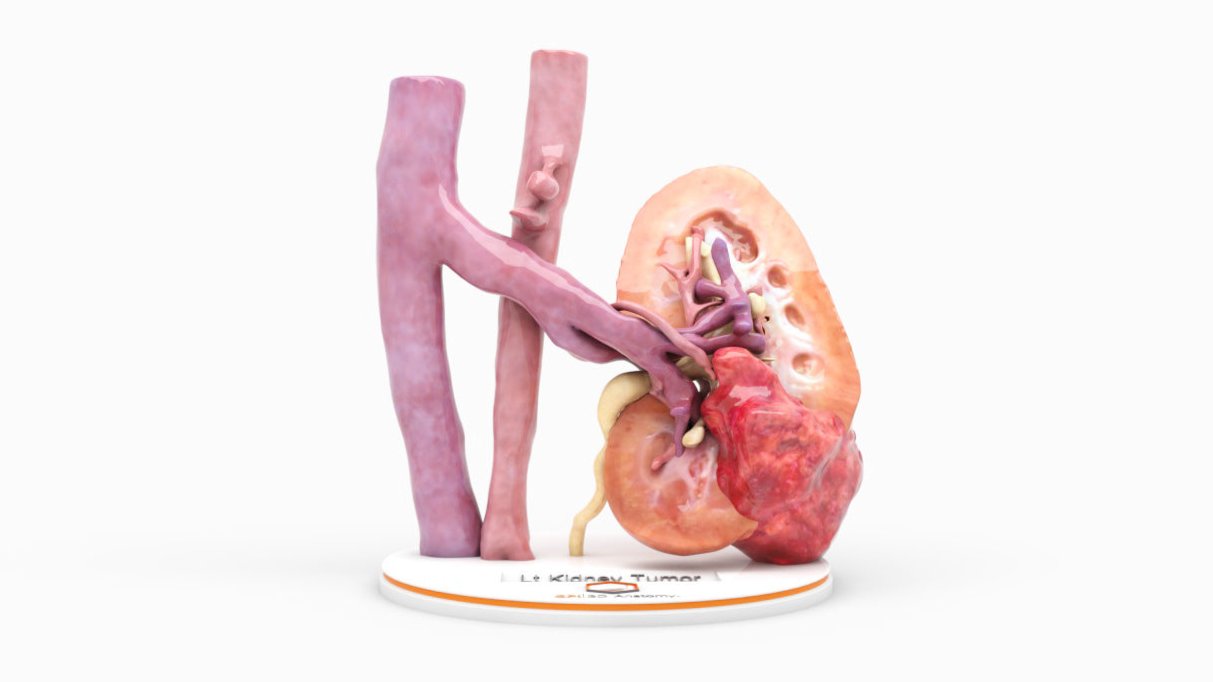
Realistic Left Kidney With Neoplasm - Male, 50 Years

$1,320.00
Shipping calculated at checkout.

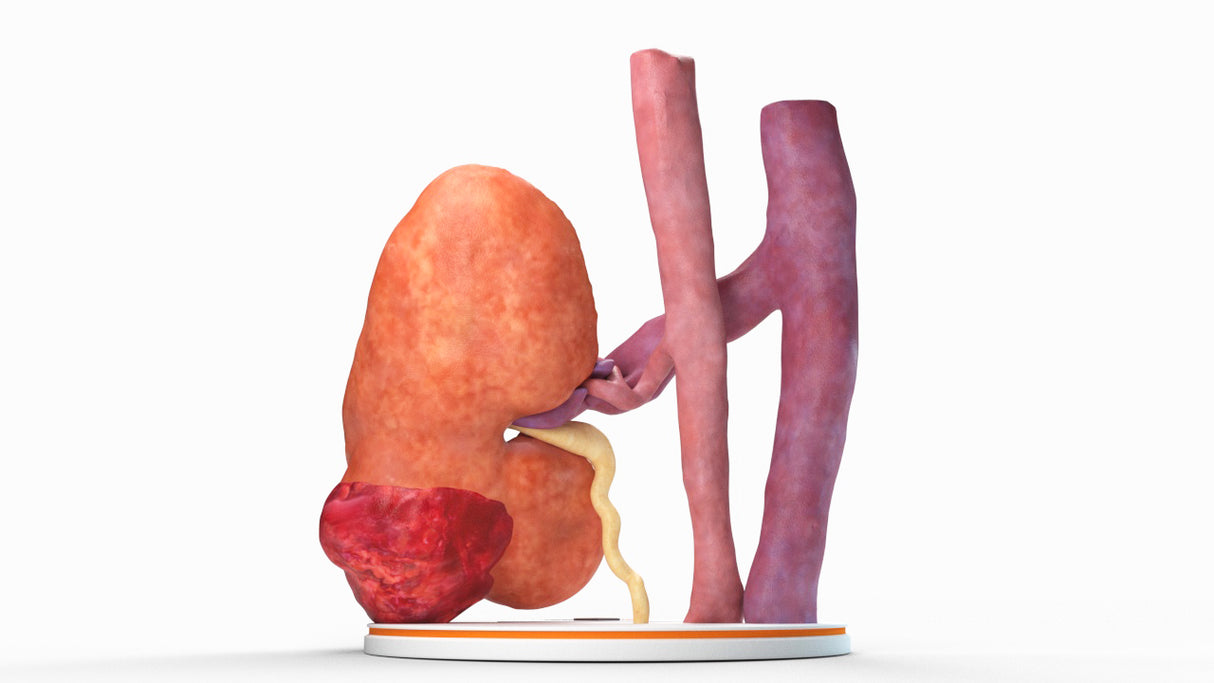
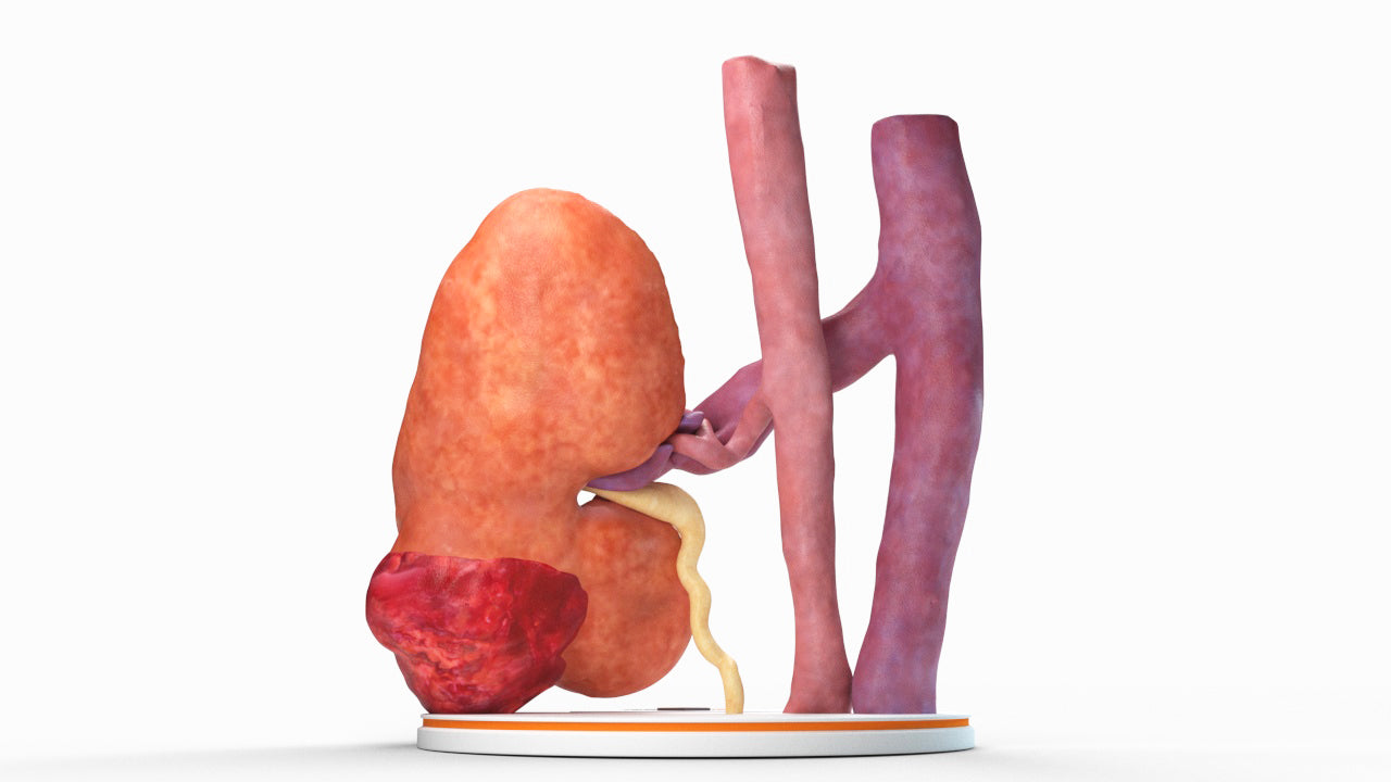
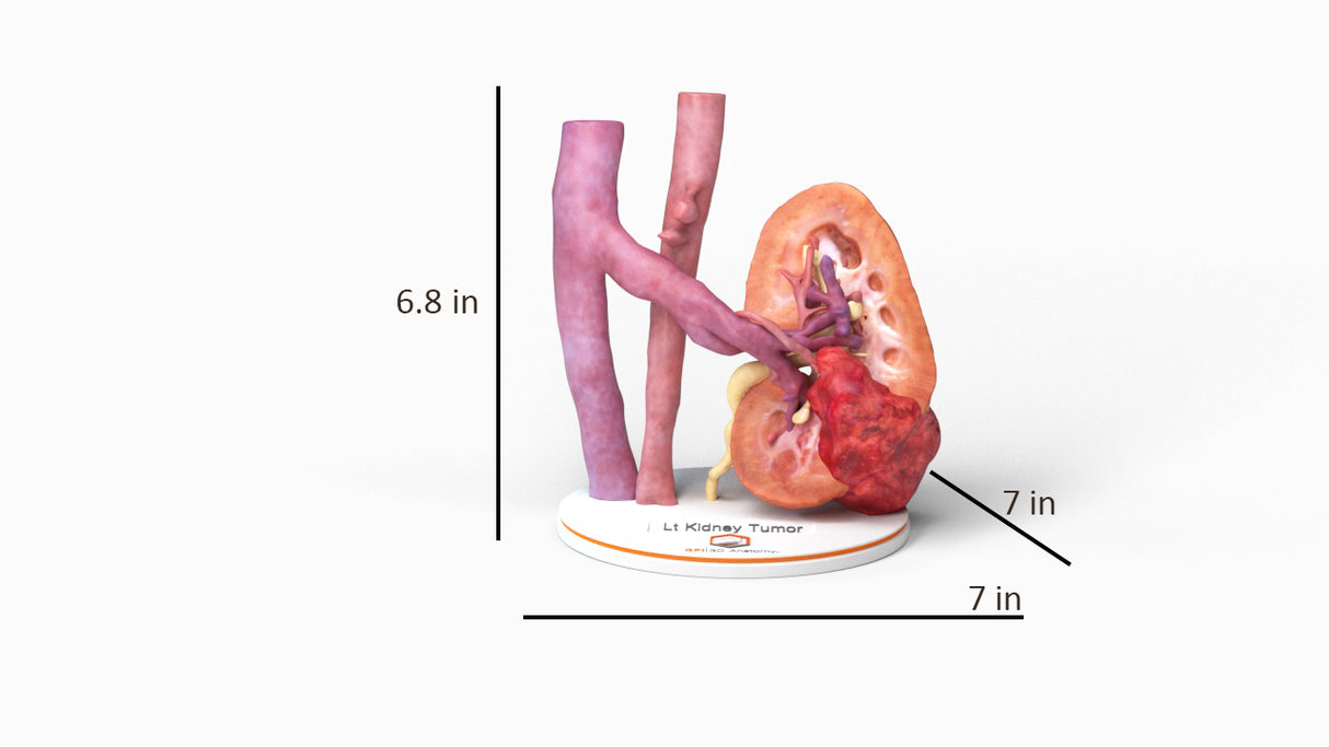
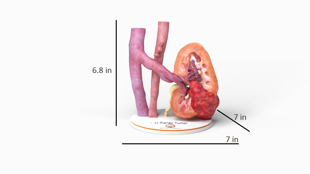
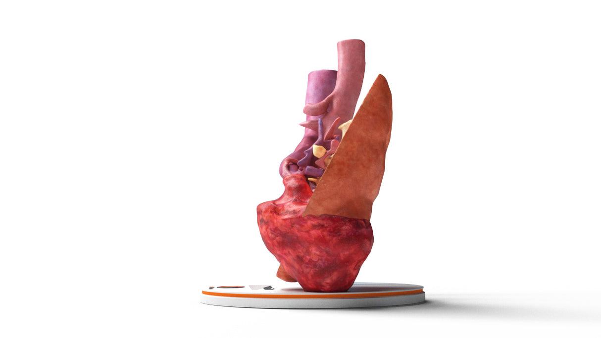
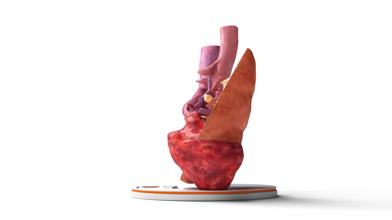
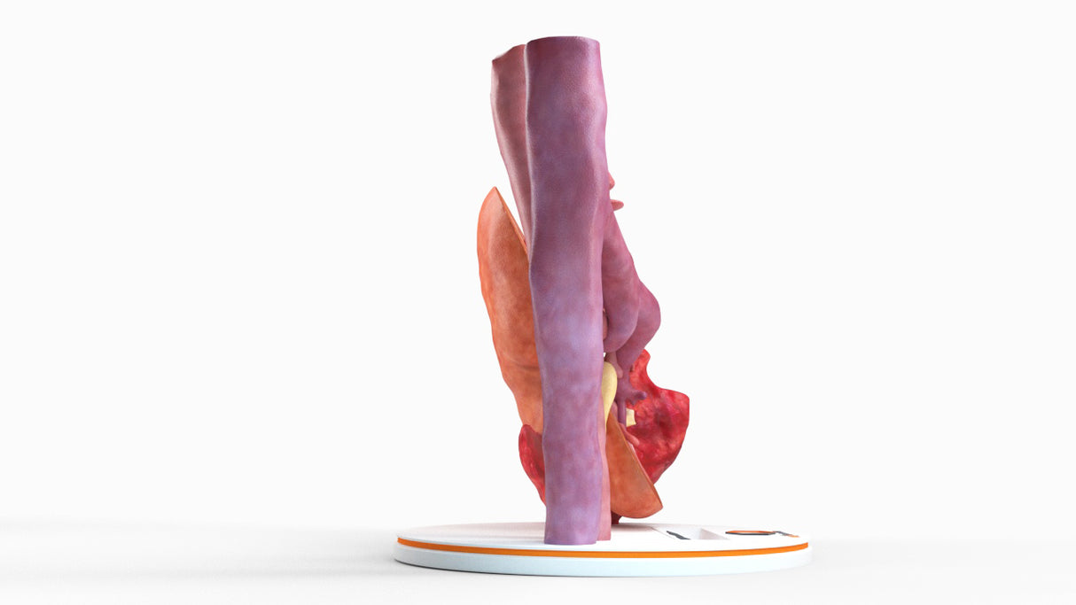
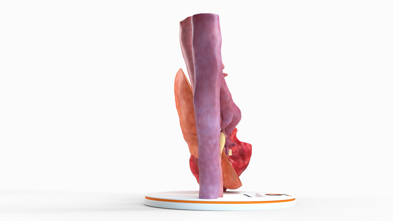
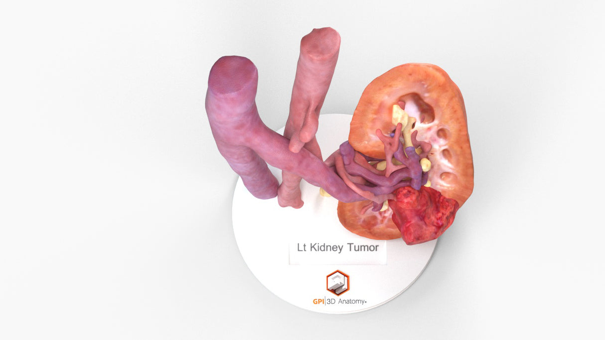
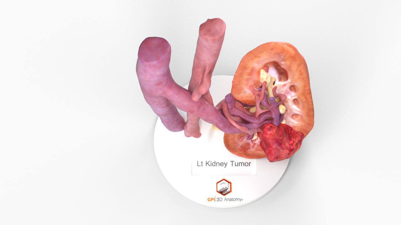
The model was created from a multiphase abdominal CT protocol including CT angiogram, Corticomedullary phase CT, CT Venogram, and delayed CT Urogram. The model depicts the interior of the renal parenchyma to reveal the inner anatomy including detailed renal artery and renal vein branches, urinary collecting systems, ureter, and their relationship to the kidney cancer (green).

Designed using real patient imaging scans and the latest 3D printing technologies, in collaboration with Mayo Clinic.

Dimensions & Features

100%: 7in x 7in x 6.8in

About the Condition

Renal Cell Cancer:
Kidney cancer results from an unregulated growth of cells in the kidney leading to the formation of a tumor. The most common tumor type, >85%, is renal cell carcinoma. Small tumors are generally asymptomatic but as the tumor grows they can present with flank pain, blood in the urine, unexplained weight loss, or a mass in the side of the abdomen. It is generally not known why kidney cancer occurs but several risk factors including obesity, smoking tobacco, high blood pressure, advanced age, and some hereditary syndromes increase a person’s risk. Prognosis is based on the stage of the tumor, which is determined by the size of the tumor, if it is contained inside the kidney, and if it has spread to adjacent organs, lymph nodes, or distant sites.

Treatment:
Surgery is the main stay of treatment.
• Partial nephrectomy, removing only the tumor and leaving the remaining healthy kidney
• Radical nephrectomy, removing the entire kidney and if needed adjacent organs and lymph nodes.

Other less invasive procedures such as cryoablation (freezing the tumor) or radiation are used in certain clinical situations.

Chemotherapy, Immunotherapy, and radiation can also be used depending on the type and stage of the cancer.

Benefits of 3D Printing

3D-printed anatomy models offer a variety of advantages for surgical planning, patient education and medical research, including:

∙ Greater accuracy and detail than traditional anatomical models. 3D-printed models are created from digital scans of a patient's anatomy, which ensures that they are as close as possible to an exact replica of real human anatomy.

∙ More versatility than traditional anatomical models. 3D-printed models can be customized to meet your specific needs, whether planning a complex surgical procedure, training with real patient data or facilitating personalized patient communication.

Not limited to standard manufacturing, 3DP provides the best opportunity to produce accurate models in natural organic shapes, sizes, and colors; creating the best representation of real human anatomy.

Why Buy With Us

  • All models are designed and produced in collaboration with Mayo Clinic 3D Anatomical Modeling Laboratories.

  • GPI Anatomicals is a leading provider of anatomical models for medical education and research, with a 40-year history of innovation and its products are used by healthcare professionals around the world.

  • READY TO SHIP  IN 1 – 2 WEEKS

    Free Shipping – Ground

    Shipping Insurance

    A Signature will be REQUIRED at time of delivery

Product comparison grid

A table comparing the facets of 2 products
Facet
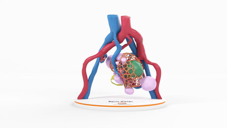
Lattice Pelvic Kidney With Neoplasm and Cysts - Male, 78 Years
View details
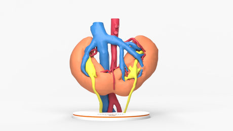
Horseshoe Kidney With Renal Cell Carcinoma (RCC) – Male, 41 Years
View details
GPI 3D Anatomy

Realistic Left Kidney With Neoplasm - Male, 50 Years

$1,320.00

The model was created from a multiphase abdominal CT protocol including CT angiogram, Corticomedullary phase CT, CT Venogram, and delayed CT Urogram. The model depicts the interior of the renal parenchyma to reveal the inner anatomy including detailed renal artery and renal vein branches, urinary collecting systems, ureter, and their relationship to the kidney cancer (green).

View product