Realistic Cervical Spine Mass - Female, 19 Years
Couldn't load pickup availability
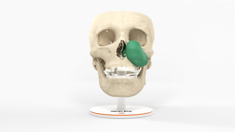
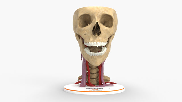
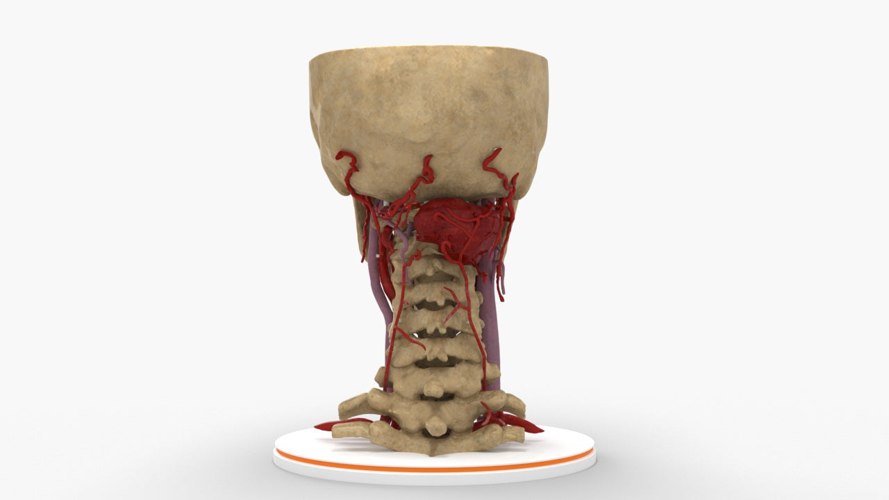
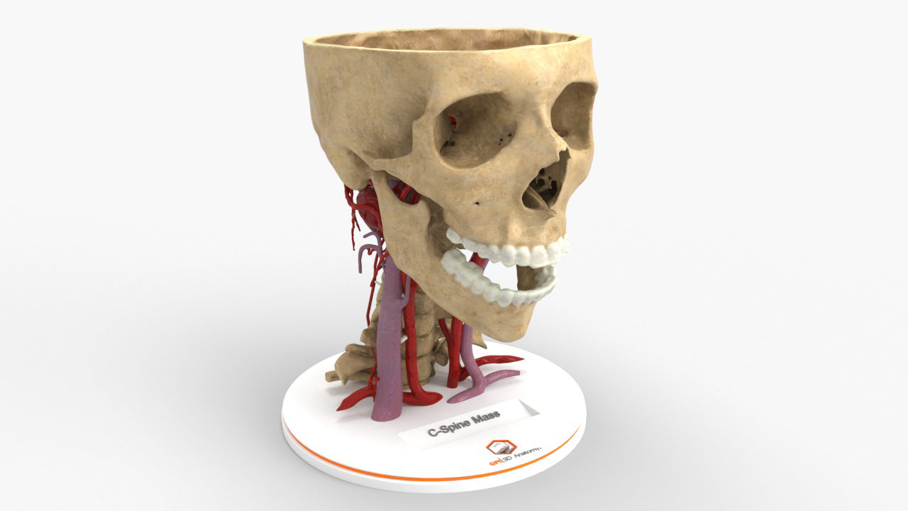
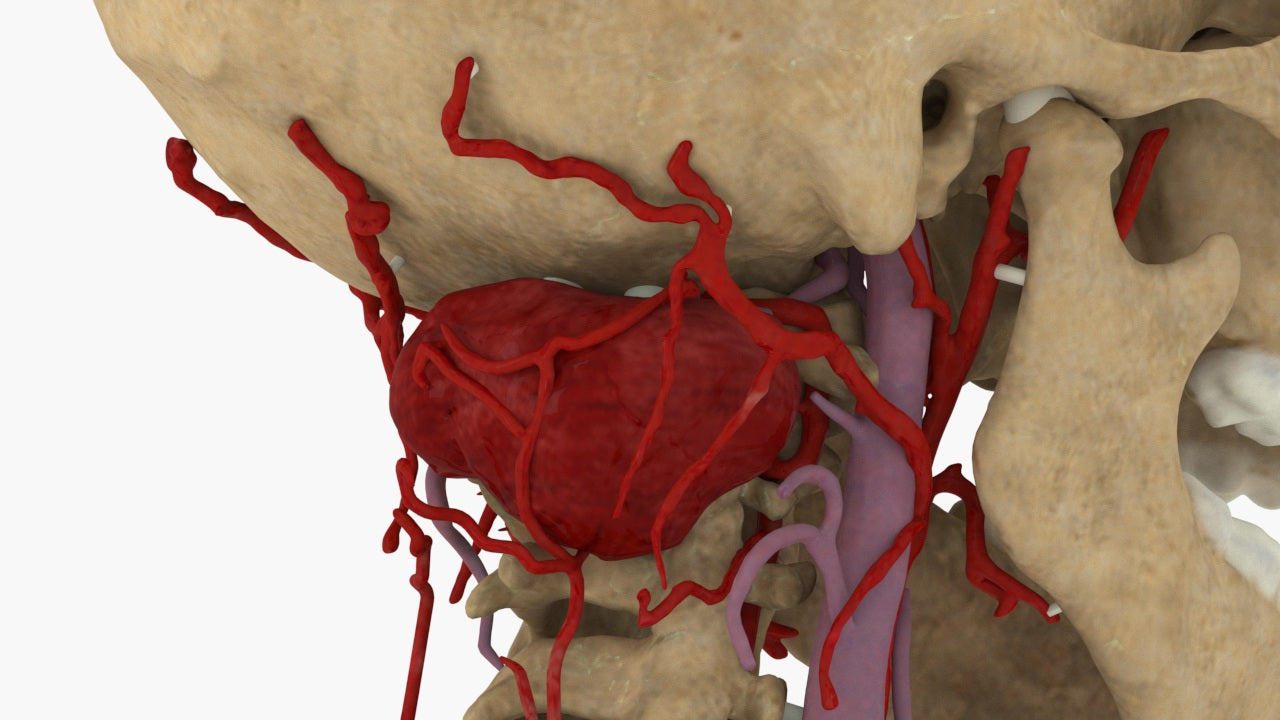
Posterior Cervical Spine Tumor: Green
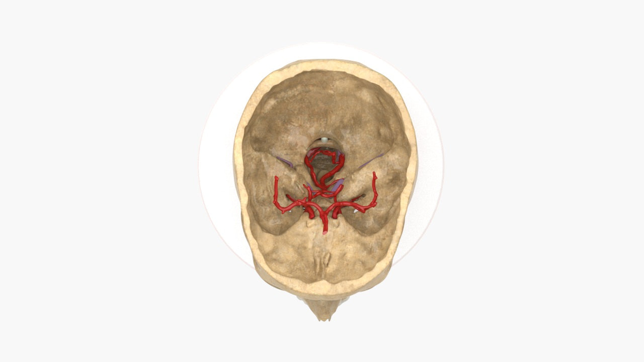
The model was created by combining MRI and CT scans of the cervical spine for preoperative planning and patient education. A realistic texture was applied to the bones and the vasculature, ending in a highly detailed representation of a sarcoma (green) located in the posterior cervical spine, aiding in a realistic 3D representation.
Designed using MRI and CT imaging scans and the latest 3D printing technologies, in collaboration with Mayo Clinic.
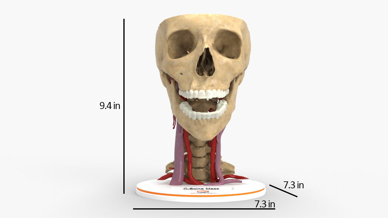
Dimensions & Features

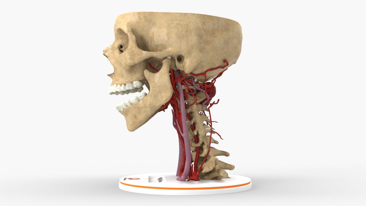
Realistic Cervical Spine Mass - Female, 19 Years
About the Condition
Benefits of 3D Printing
3D-printed anatomy models offer a variety of advantages for surgical planning, patient education and medical research, including:
∙ Greater accuracy and detail than traditional anatomical models. 3D-printed models are created from digital scans of a patient's anatomy, which ensures that they are as close as possible to an exact replica of real human anatomy.
∙ More versatility than traditional anatomical models. 3D-printed models can be customized to meet your specific needs, whether planning a complex surgical procedure, training with real patient data or facilitating personalized patient communication.
Not limited to standard manufacturing, 3DP provides the best opportunity to produce accurate models in natural organic shapes, sizes, and colors; creating the best representation of real human anatomy.
Why Buy With Us
Product comparison grid
| Facet |
|---|