About the Condition:
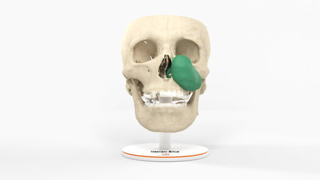
The paranasal sinuses are air-filled cavities located within the bones of the skull and face. The maxillary sinus is the largest of all the sinuses, with direct communication to the maxillary teeth and nasal cavity. Tumors involving the maxillary sinus can be benign (noncancerous) or malignant (cancerous). 70% of all paranasal tumors originate in the maxillary sinus, given its size and cellular make-up. 80% of all primary malignant tumors in the maxillary sinus are squamous cell carcinomas arising from the mucosal lining like this tumor. Enlarging tumors may present due to symptoms related to obstruction of the drainage pathway into the nose or direct pressure on surrounding anatomy like the orbit. These include difficulty breathing through the nose, sinus pressure, headache, teeth pain, double vision, facial swelling, or watery eyes. Maxillary sinus cancers often grow undetected until they are quite large due to the sinus's open space, allowing tumors to expand significantly before being discovered. As they progress, they may invade nearby structures, including the orbit and palate, requiring aggressive surgical treatment and adjuvant radiation and/or chemotherapy.
Risk factors that increase the risk of nasal and paranasal tumors include smoking tobacco, chronic exposure to air pollution, chemicals, and irritants, including wood dust, fumes from glue, rubbing alcohol, formaldehyde, dust from flour, chromium, and nickel. Being exposed to human papillomavirus, also called HPV, a common virus that's passed through sexual contact, is another risk factor.
Benign Tumors: Papillomas (Inverted and Fungiform Papillomas), Osteomas, Fibrous Dysplasia, Angiofibromas.
Malignant Tumors: Squamous Cell Carcinoma, Adenocarcinoma, Mucoepidermoid Carcinoma, Adenoid Cystic Carcinoma, sinonasal undifferentiated carcinoma (SNUC), , lymphoma, Melanoma, Osteosarcoma, Chondrosarcoma, Rhabdomyosarcoma.
Treatments for malignant cancers of the maxillary sinus
The prognosis and treatment options depend on where the tumor is in the paranasal sinus and whether it has spread outside the sinus. The size of the cancer, the type of tumor, and the patient's age and general health are all used to determine the best treatment plan. A doctor will perform a physical exam and then typically examine the nasal passageways and sinus with nasal endoscopy. If a lesion is seen, a biopsy is typically performed, and then a CT and/or MRI scan is performed to determine the extent of the tumor and spread outside of the sinus.
For malignant tumors, surgery is often the primary approach, involving the removal of the tumor and affected structures. Several factors determine the aggressiveness of the surgery. In advanced cases, a maxillectomy may be required, and a vascularized free flap may be used to reconstruct the midface. Radiation therapy is commonly used post-surgery to eliminate any remaining cancer cells or as the primary treatment for inoperable tumors. Chemotherapy may be combined with radiation and surgery for more aggressive or metastatic cases. Targeted therapies and immunotherapy are emerging as potential options for specific cancer types. Early detection and a multidisciplinary approach including oncologists, radiation oncologists, surgeons, and radiologists all improve treatment outcomes.
Maxillary Sinus Anatomy:
The maxillary sinus is the most extensive paranasal sinuses within the maxilla (upper jaw). It is a pyramid-like, air-filled space that plays a role in humidifying and filtering inhaled air and contributes to voice resonance. Its proximity to the upper teeth makes it clinically significant, as infections or dental procedures can sometimes affect the sinus, leading to sinusitis or oroantral fistulas. The maxillary sinus drains into the nasal cavity through the ostium, an opening in the middle meatus. The maxillary sinus is unique because it does not drain by gravity but relies on mucociliary action to move mucus upward, making it prone to congestion and infection if obstructed.