Chest Wall with Tumor

$1,780.00
Shipping calculated at checkout.

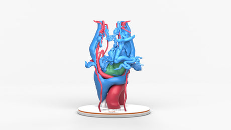
Chondrosarcoma of the chest wall

Model was created by combining a MRI co-registered to a CT scan of the chest wall for patient education and preoperative/reconstructive planning.

Designed using real patient imaging scans and the latest 3D printing technologies, in collaboration with Mayo Clinic.

Dimensions & Features

100%: 8in x 10in x 12.5in

About the Condition

Chondrosarcoma:
A rare malignant tumor that begins in the bone and is caused by unregulated growth of cartilage forming cells. Although chondrosarcomas are more common in the pelvis they are the most common primary malignant tumor of the chest wall. Most patients present with an enlarging, painful anterior chest wall mass arising from the junction between the cartilage and rib or sternum.

Chest Wall:
A multilayered framework of the thorax which facilitates respiratory function and serves to protect the heart, lungs, and major blood vessels. It consists of the sternum, ribs, cartilage, fascia, pleura, spine, and muscles. Both primary and secondary tumors can affect the chest wall. A primary tumor arises in one of the tissues of the chest wall and grows into the adjacent structures. A secondary tumor either directly invades the chest wall from adjacent organs (lung or breast) or from metastatic spread of another tumor from a distant site.

Treatment:
Primary chest wall tumors are rare affecting < 2% of the population. 50-80% are malignant and the remainder benign. Surgical resection is the first line of treatment depending on size, invasiveness, preexisting health conditions, and whether there is metastasis. Depending on the type and grade of the tumor the patient may need additional chemotherapy or radiation. If the resected lesion is large the chest wall may need to be reconstructed to minimize the effect on respiratory function.

Benefits of 3D Printing

3D-printed anatomy models offer a variety of advantages for surgical planning, patient education and medical research, including:

∙ Greater accuracy and detail than traditional anatomical models. 3D-printed models are created from digital scans of a patient's anatomy, which ensures that they are as close as possible to an exact replica of real human anatomy.

∙ More versatility than traditional anatomical models. 3D-printed models can be customized to meet your specific needs, whether planning a complex surgical procedure, training with real patient data or facilitating personalized patient communication.

Not limited to standard manufacturing, 3DP provides the best opportunity to produce accurate models in natural organic shapes, sizes, and colors; creating the best representation of real human anatomy.

Why Buy With Us

  • All models are designed and produced in collaboration with Mayo Clinic 3D Anatomical Modeling Laboratories.

  • GPI Anatomicals is a leading provider of anatomical models for medical education and research, with a 40-year history of innovation and its products are used by healthcare professionals around the world.

  • READY TO SHIP  IN 1 – 2 WEEKS

    Free Shipping – Ground

    Shipping Insurance

    A Signature will be REQUIRED at time of delivery

Product comparison grid

A table comparing the facets of 3 products
Facet
Chest Wall with Tumor
View details
Chest With Mediastinal Paraganglioma – Male, 50 Years
View details
Tracheobronchial Tree With Lymph Nodes - Male, 28 Years
View details
GPI 3D Anatomy

Chest Wall with Tumor

$1,780.00

Chondrosarcoma of the chest wall

Model was created by combining a MRI co-registered to a CT scan of the chest wall for patient education and preoperative/reconstructive planning.

View product