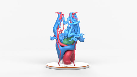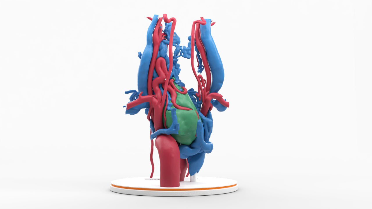
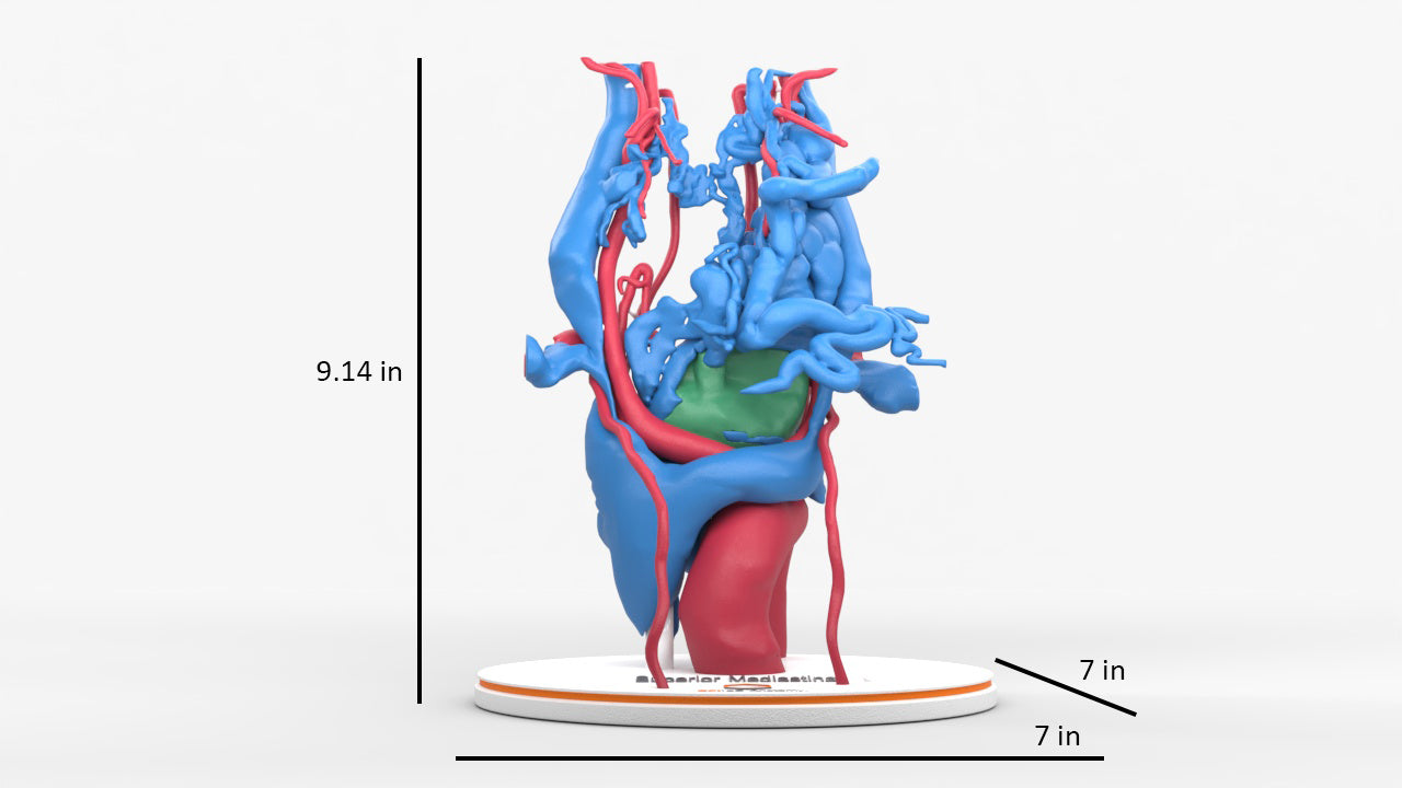
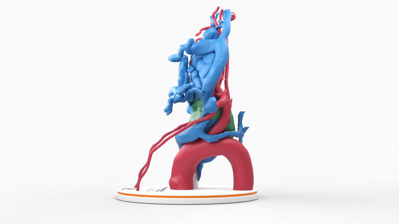
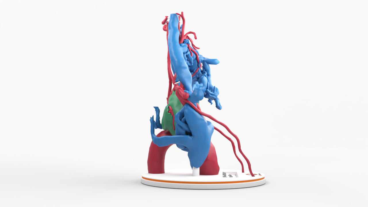
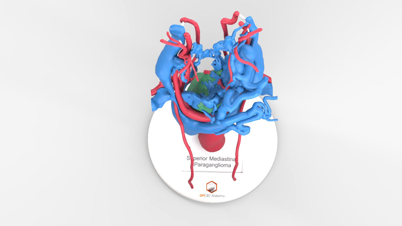
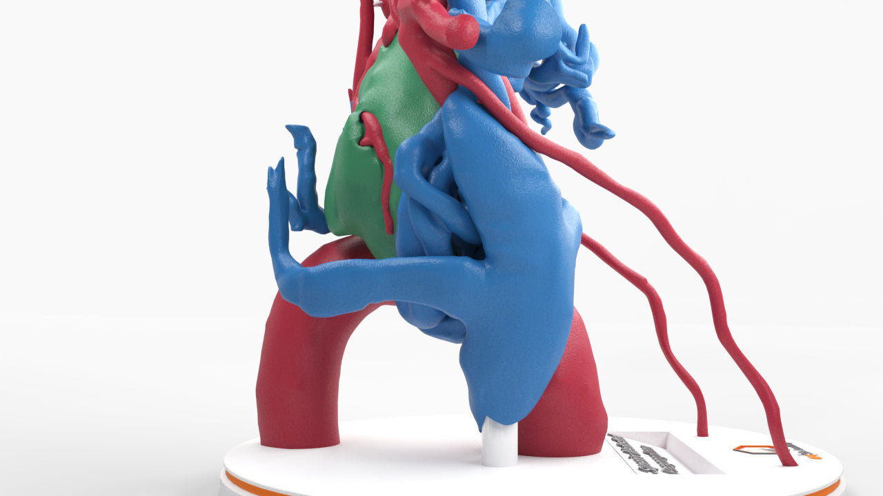
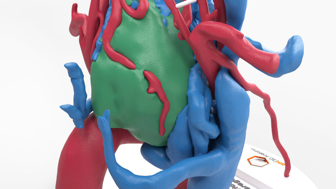
Chest With Mediastinal Paraganglioma – Male, 50 Years
Pricing and Availability Upon Request
Contact us: sales@gpianatomy.com
Chest with middle mediastinal paraganglioma, a rare tumor type derived from chromaffin cells (neuroectodermal cells), with abnormal increased arterial vascularity supplying the tumor and multiple enlarged draining veins. Due to the increased vascularity, this tumor type is at high risk of massive blood loss intraoperatively. Detailed surgical technique and preoperative blood pressure control are essential in the prevention and management of complications.
Complete surgical resection remains the standard of care. Prognosis after complete resection is favorable.
Designed using MRI and CT imaging scans and the latest 3D printing technologies, in collaboration with Mayo Clinic.
Dimensions & Features

Chest With Mediastinal Paraganglioma – Male, 50 Years
About the Condition
Benefits of 3D Printing
3D-printed anatomy models offer a variety of advantages for surgical planning, patient education and medical research, including:
∙ Greater accuracy and detail than traditional anatomical models. 3D-printed models are created from digital scans of a patient's anatomy, which ensures that they are as close as possible to an exact replica of real human anatomy.
∙ More versatility than traditional anatomical models. 3D-printed models can be customized to meet your specific needs, whether planning a complex surgical procedure, training with real patient data or facilitating personalized patient communication.
Not limited to standard manufacturing, 3DP provides the best opportunity to produce accurate models in natural organic shapes, sizes, and colors; creating the best representation of real human anatomy.
Why Buy With Us
Product comparison grid
| Facet |
|---|