Pelvic Hernia – Female, 48 Years
Couldn't load pickup availability
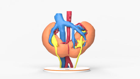
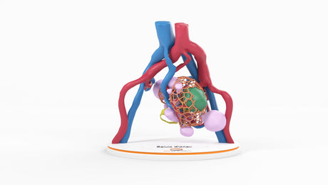
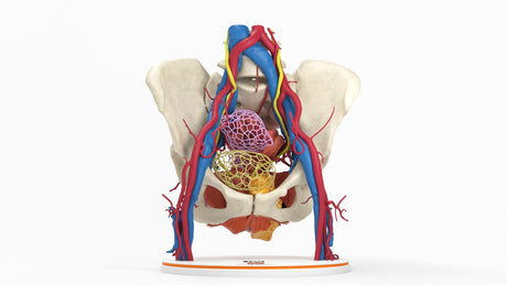
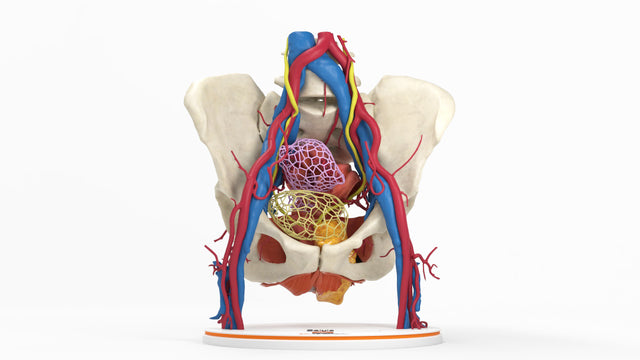
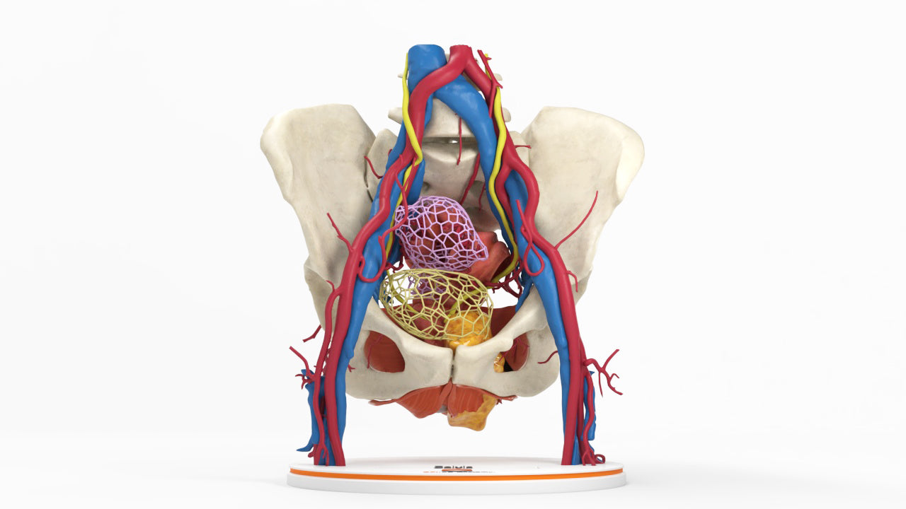
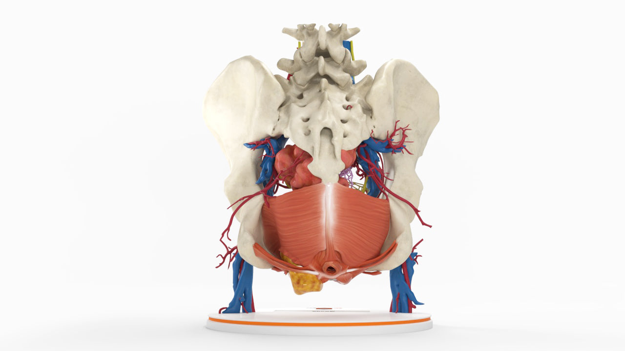
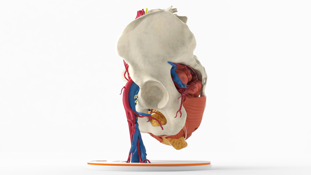
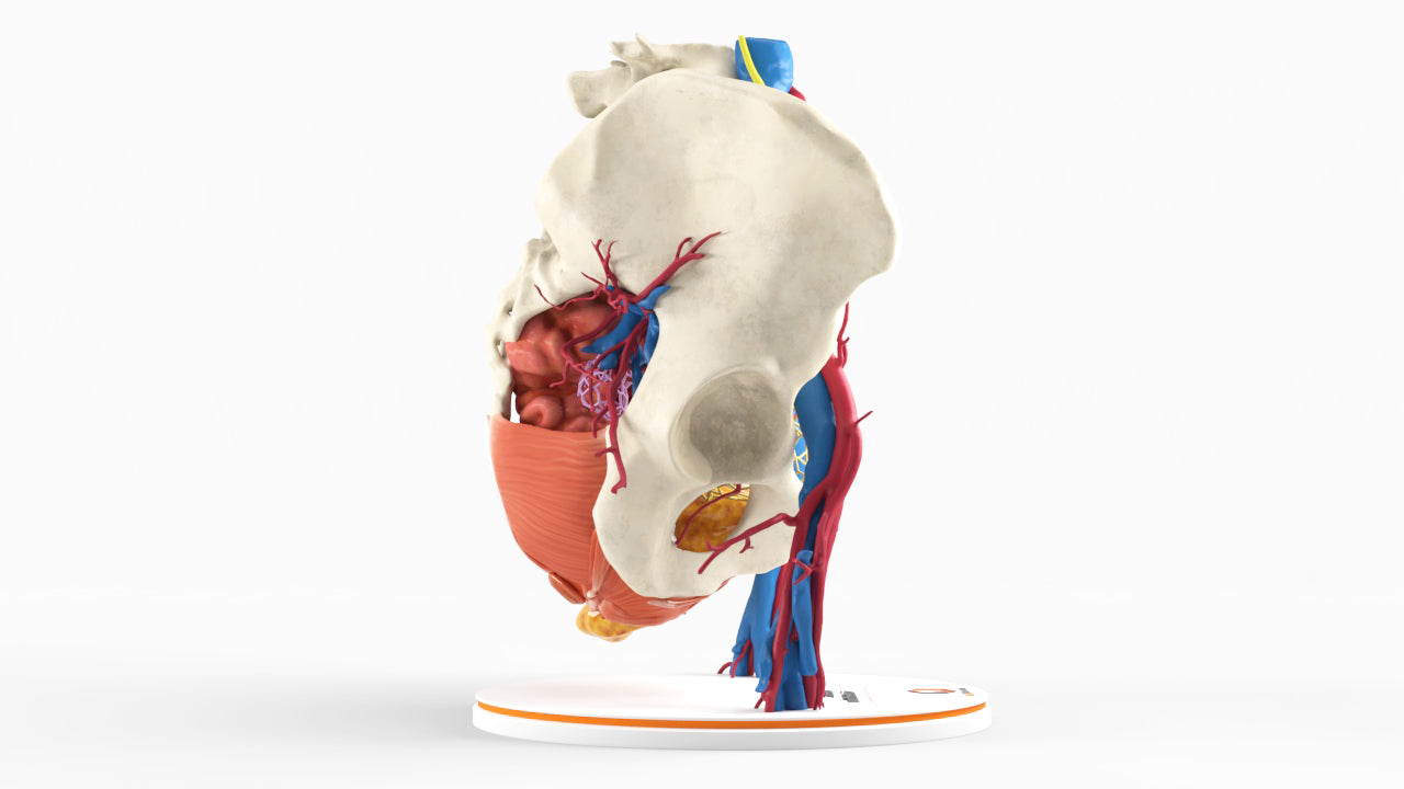
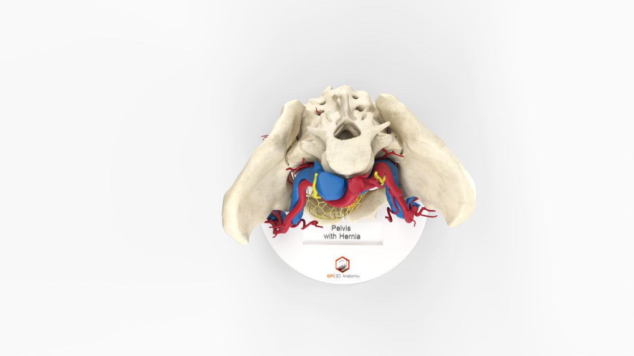
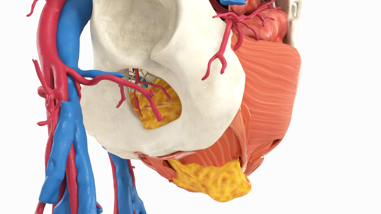
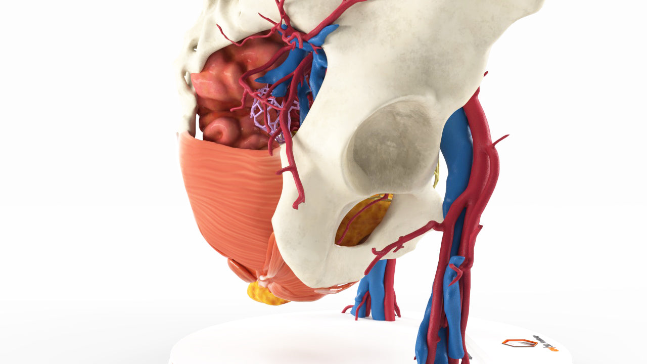
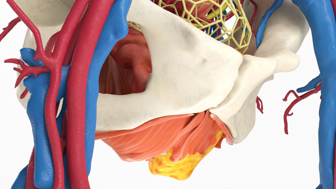
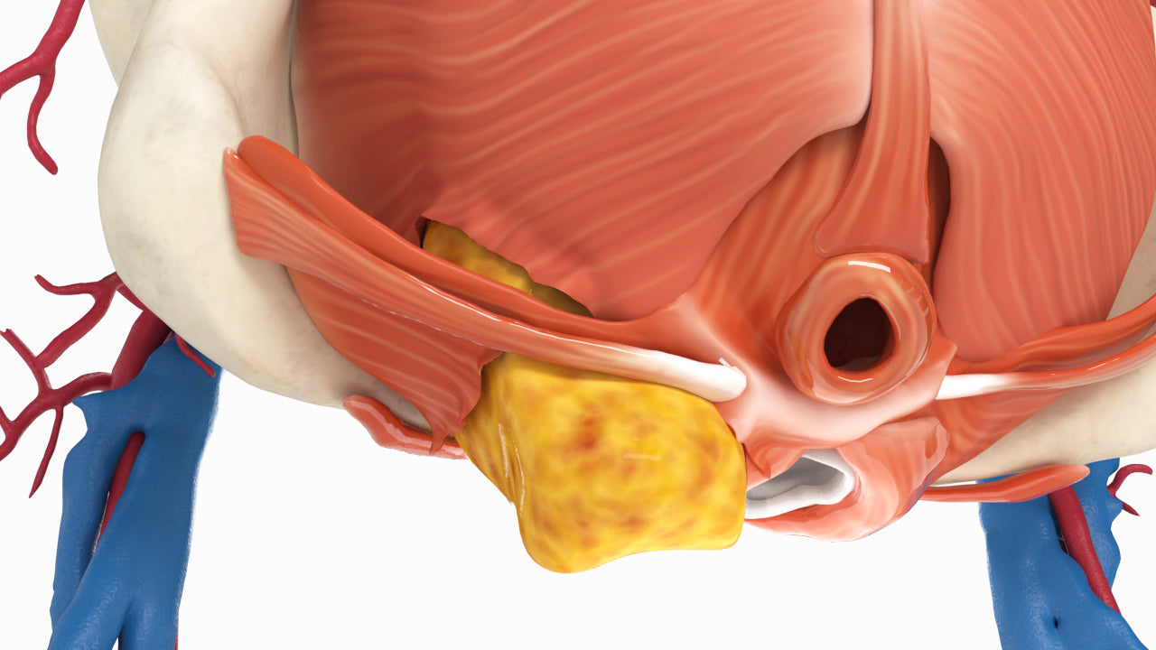
Perineal hernia with pelvic floor dysfunction, a rare but known complication after pelvic surgery. Includes lattice bladder, uterus, vagina, rectum and pelvic floor muscles supported by vasculature.
Surgical repair including transabdominal, transperineal and combined approaches is the definitive treatment and may include primary closure of the defect and/or reinforcement of the pelvic floor using mesh.
Designed using MRI and CT imaging scans and the latest 3D printing technologies, in collaboration with Mayo Clinic.
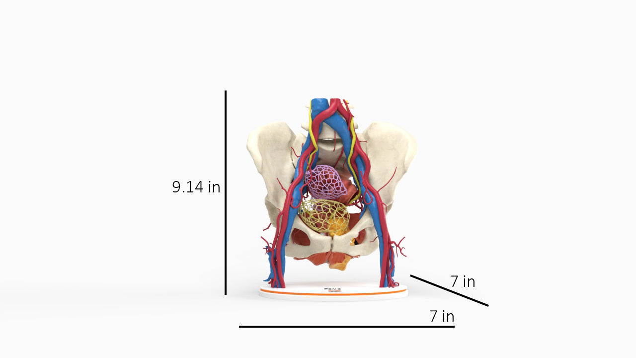
Dimensions & Features

Pelvic Hernia – Female, 48 Years
About the Condition
Benefits of 3D Printing
3D-printed anatomy models offer a variety of advantages for surgical planning, patient education and medical research, including:
∙ Greater accuracy and detail than traditional anatomical models. 3D-printed models are created from digital scans of a patient's anatomy, which ensures that they are as close as possible to an exact replica of real human anatomy.
∙ More versatility than traditional anatomical models. 3D-printed models can be customized to meet your specific needs, whether planning a complex surgical procedure, training with real patient data or facilitating personalized patient communication.
Not limited to standard manufacturing, 3DP provides the best opportunity to produce accurate models in natural organic shapes, sizes, and colors; creating the best representation of real human anatomy.
Why Buy With Us
Product comparison grid
| Facet |
|---|