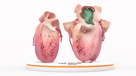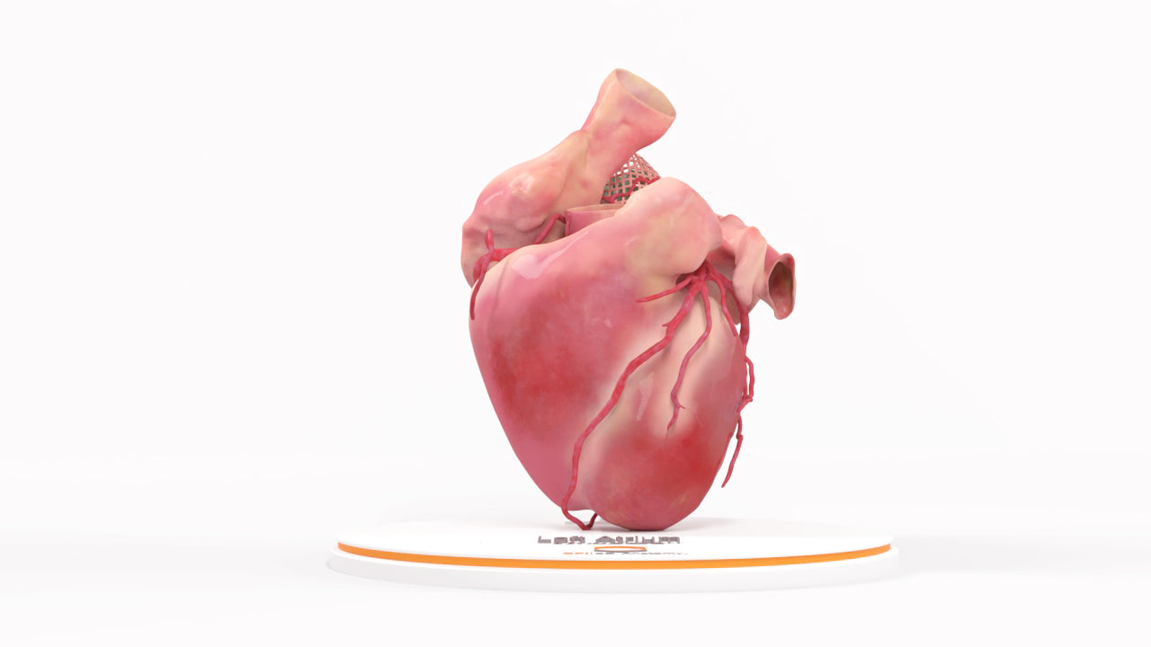
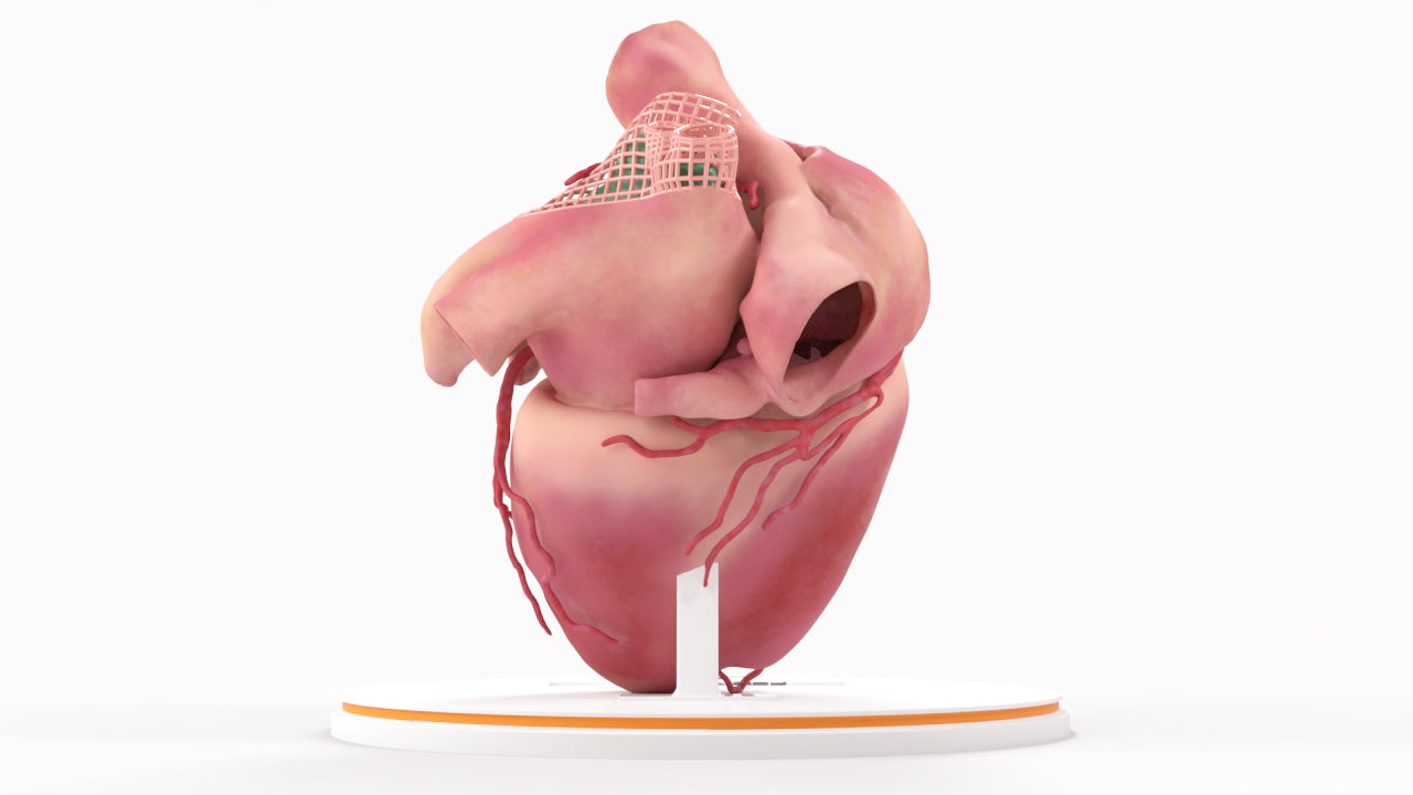
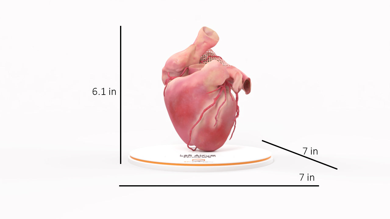
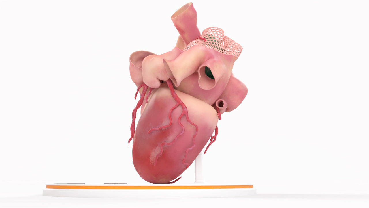
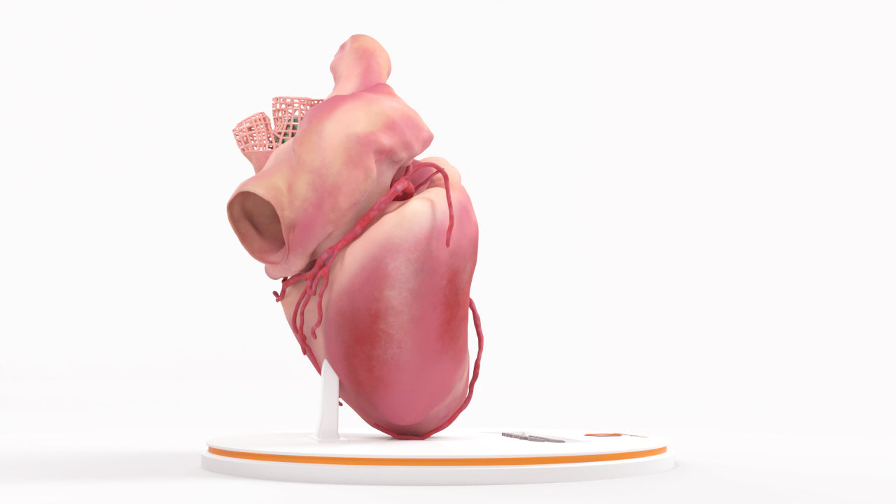
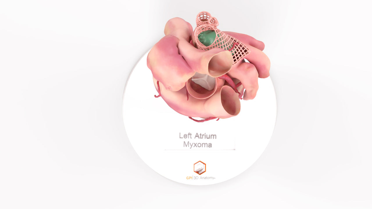
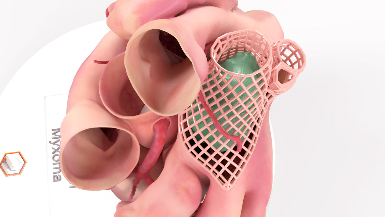
Left Atrium Myxoma, Closed - Female, 73 Years
Couldn't load pickup availability
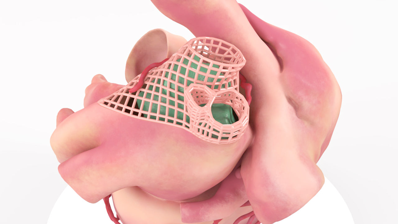
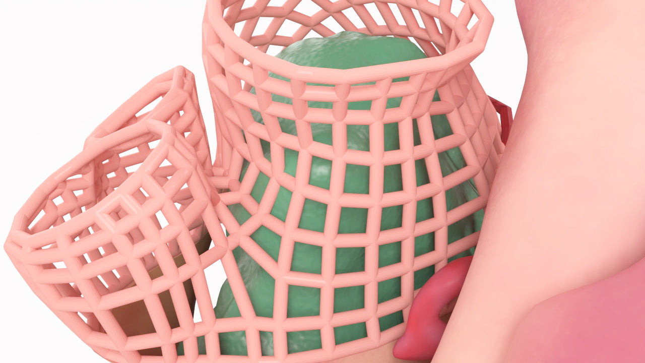
Left Atrial Myxoma in a 73 year old female: Closed
Model was created by co-registering a cardiac MRI to a gated CT Angiogram of the heart and segmenting the critical anatomy and pathology for preoperative planning and patient education. A lattice design was added to the pulmonary veins and superior left atrium to allow visualization of the tumor.
Designed using MRI and CT imaging scans and the latest 3D printing technologies, in collaboration with Mayo Clinic.
Dimensions & Features
Left Atrium Myxoma, Closed - Female, 73 Years
About the Condition
Benefits of 3D Printing
3D-printed anatomy models offer a variety of advantages for surgical planning, patient education and medical research, including:
∙ Greater accuracy and detail than traditional anatomical models. 3D-printed models are created from digital scans of a patient's anatomy, which ensures that they are as close as possible to an exact replica of real human anatomy.
∙ More versatility than traditional anatomical models. 3D-printed models can be customized to meet your specific needs, whether planning a complex surgical procedure, training with real patient data or facilitating personalized patient communication.
Not limited to standard manufacturing, 3DP provides the best opportunity to produce accurate models in natural organic shapes, sizes, and colors; creating the best representation of real human anatomy.
Why Buy With Us
Product comparison grid
| Facet |
|---|