The abdominal and pelvic vasculature consists of several branching arterial systems, systemic venous tributaries, and portal venous systems.
Arterial System:
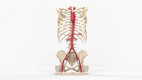
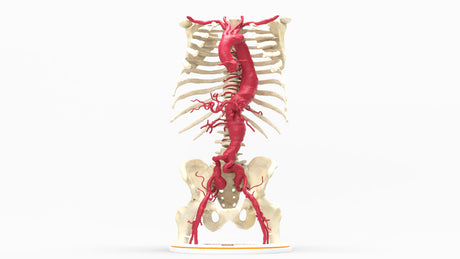
The abdominal aorta is the continuation of the thoracic aorta and extends from the aortic hiatus (T12) to the level of the fourth lumbar level, where it bifurcates into the right and left common iliac arteries. There are 3 main unpaired visceral branches supplying end organs. First is the celiac trunk, which trifurcates into the left gastric artery, common hepatic artery, and splenic artery supplying the liver, stomach, and spleen. Second, the superior mesenteric artery supplies blood to the small intestine, pancreas, cecum, and ascending and proximal transverse colon. Lastly, the inferior mesenteric artery supplies blood to the remaining parts of the colon and rectum. Several paired visceral branches include the suprarenal arteries supplying the adrenal glands, the renal arteries supplying the kidneys, and the gonadal arteries supplying the gonadal structures. The remaining branches include the inferior phrenic arteries, the five lumbar arteries, and the middle sacral artery supplying oxygenated blood to the diaphragm, lumbar spine, sacrum, and coccyx.
The pelvic arterial systems are not shown in great detail in this model. The Iliac arteries bifurcate into the internal and external iliac arteries. The external iliac artery gives rise to the inferior epigastric and deep circumflex arteries and continues as the femoral arteries.
Venous System:
The venous drainage of the abdominal and pelvic regions involves the systemic venous system through the inferior vena cava (IVC) and the portal venous system (PVS) largely through the portal vein.
Systemic Venous System:
The systemic venous system is responsible for returning deoxygenated blood from the body to the heart, where the lungs can reoxygenate it before recirculating through the body. The abdomen and pelvis consist of a vast network of veins that collect blood from the lower extremities, abdominal and pelvic organs, and visceral structures, ultimately draining into the inferior vena cava (IVC), which drains directly into the right atrium. The IVC is formed by the union of the common iliac veins at the level of the fifth lumbar vertebra ascending to the right of midline, anterior to the vertebral column, in the retroperitoneal space, finally traversing the tendinous portion of the diaphragm at the level of the eighth thoracic vertebra. There are several tributaries to the IVC during its ascent through the abdomen, including the paired renal veins draining the kidneys, the hepatic veins draining blood from the liver, the suprarenal veins draining the adrenals, the paired gonadal veins, the inferior phrenic veins, the lumbar veins.
Portal Venous System:
The portal venous system (PVS) drains blood from the gastrointestinal tract, gallbladder, pancreas, and spleen to the liver to process nutrients and filtrate toxins. The Portal Vein (PV), formed from the confluence of the superior mesenteric and splenic veins, is the PVS's main blood, carrying approximately 75% of the blood flow to the liver. Having a dual blood supply, the liver also receives 25% of its blood from the hepatic arteries. The PVS also includes the inferior mesenteric vein, which most commonly drains into the splenic vein. The Portal vein drains into the sinusoids, specialized capillary-like vessels, and mixes with the hepatic arterial blood supply. Due to the sinusoid lacking a basement membrane, there is a direct exchange between the sinusoids and the hepatocytes, as well as specialized cells called Kupffer cells, before entering the central vein in the hepatic lobule. The central veins drain into the hepatic veins and inferior vena cava, which begins the systemic venous system.
Common Pathologies:
Arterial: Aortic aneurysms, Aortic dissections, mesenteric ischemia, and renal artery stenosis.
Venous: Portal venous hypertension, portal vein thrombosis, nutcracker syndrome (compression of the left renal vein between the aorta and the superior mesenteric artery)